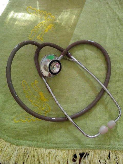
A stethoscope is a medical device for listening to sounds inside the body. The initial stethoscope was invented in the early 19th century by French physician Ren� Laennec, but was actually trying to achieve a rather different end: doctor-patient distance....
Wednesday, June 24, 2015
Surgical Risk Factors And Preventive Strategies : Perioperative Nursing
Obesity
Poor Nutrition
Fluid and Electrolyte Imbalance
Aging
Presence of Cardiovascular Disease
Therapeutic Approach
Presence of Pulmonary and Upper Respiratory Disease
Concurrent or Prior Pharmacotherapy
Read More
Danger
-
Increases the difficulty involved in the technical aspects of performing surgery; risk for wound dehiscence is greater
-
Increases the likelihood of infection because of compromised tissue perfusion
-
Increases the potential for postoperative pneumonia and other pulmonary complications because obese patients chronically hypoventilate
-
Increases demands on the heart, leading to cardiovascular compromise
-
Increases the risk for airway complications
-
Alters the response to many drugs and anesthetics
-
Decreases the likelihood of early ambulation
Therapeutic Approach
-
Encourage weight reduction if time permits.
-
Anticipate postoperative obesity-related complications.
-
Be extremely vigilant for respiratory complications.
-
Carefully splint abdominal incisions when moving or coughing.
-
Be aware that some drugs should be dosed according to ideal body weight versus actual weight (owing to fat content); otherwise, an overdose may occur (digoxin [Lanoxin], lidocaine [Xylocaine], aminoglycosides, and theophylline [Theo-Dur]).
-
Avoid intramuscular injections in morbidly obese individuals (I.V. or subcutaneous routes preferred).
-
Never attempt to move an impaired patient without assistance or without using proper body mechanics.
-
Obtain a dietary consultation early in the patient's postoperative course.
Danger
-
Greatly impairs wound healing (especially protein and calorie deficits and a negative nitrogen balance)
-
Increases the risk of infection
Therapeutic Approach
-
Any recent (within 4 to 6 weeks) weight loss of 10% of the patient's normal body weight or decreased serum albumin should alert the health care staff to poor nutritional status and the need to investigate as to the cause of the weight loss.
-
Attempt to improve nutritional status before and after surgery. Unless contraindicated, provide a diet high in proteins, calories, and vitamins (especially vitamins C and A); this may require enteral and parenteral feeding.
-
Review a serum prealbumin level to determine recent nutritional status.
-
Recommend repair of dental caries and proper mouth hygiene to prevent infection.
Fluid and Electrolyte Imbalance
Danger
Can have adverse effects in terms of general anesthesia and the
anticipated volume losses associated with surgery, causing shock and cardiac
dysrhythmias
Patients undergoing major abdominal operations
(such as colectomies and aortic repairs) often experience a massive fluid shift
into tissues around the operative site in the form of edema (as much as 1 L or
more may be lost from circulation). Watch for the fluid shift to reverse (from
tissue to circulation) around the third postoperative day. Patients with heart
disease may develop failure due to the excess fluid “load.”
Therapeutic Approach
-
Assess the patient's fluid and electrolyte status.
-
Rehydrate the patient parenterally and orally as prescribed.
-
Monitor for evidence of electrolyte imbalance, especially Na+, K+, Mg++, Ca++.
-
Be aware of expected drainage amounts and composition; report excess and abnormalities.
-
Monitor the patient's intake and output; be sure to include all body fluid losses.
Danger
-
Potential for injury is greater in older people
-
Be aware that the cumulative effect of medications is greater in the older patient
-
Medications in the usual dosages, such as morphine, may cause confusion, disorientation, and respiratory depression
Therapeutic Approach
-
Consider using lesser doses for desired effect.
-
Anticipate problems from chronic disorders such as anemia, obesity, diabetes, hypoproteinemia.
-
Adjust nutritional intake to conform to higher protein and vitamin needs.
-
When possible, cater to set patterns in older patients, such as sleeping and eating.
Danger
-
May compound the stress of anesthesia and the operative procedure
-
May result in impaired oxygenation, cardiac rhythm, cardiac output, and circulation
-
May also produce cardiac decompensation, sudden arrhythmia, thromboembolism, acute myocardial infarction (MI), or cardiac arrest
Therapeutic Approach
-
Frequently assess heart rate and blood pressure (BP) and hemodynamic status and cardiac rhythm if indicated.
-
Avoid fluid overload (oral, parenteral, blood products) because of possible MI, angina, heart failure, and pulmonary edema.
-
Prevent prolonged immobilization, which results in venous stasis. Monitor for potential deep vein thrombosis (DVT) or pulmonary embolus.
-
Encourage position changes but avoid sudden exertion.
-
Use antiembolism stockings and/or sequential compression device intraoperatively and postoperatively.
-
Note evidence of hypoxia and initiate therapy.
Presence of Diabetes Mellitus
Danger
-
Hypoglycemia may result from food and fluid restrictions and anesthesia.
-
Hyperglycemia and ketoacidosis may be potentiated by increased catecholamines and glucocorticoids due to surgical stress.
-
Chronic hyperglycemia results in poor wound healing and susceptibility to infection.
-
Recognize the signs and symptoms of ketoacidosis and hypoglycemia, which can threaten an otherwise uneventful surgical experience. Dehydration also threatens renal function.
-
Monitor blood glucose and be prepared to administer insulin, as directed, or treat hypoglycemia.
-
Confirm what medications the patient has taken and what has been held. Facility protocol and provider preference varies, but the goal is to prevent hypoglycemia. If the patient is NPO, oral agents are usually withheld and insulin may be ordered at 75% of the usual dose.
-
Reassure the diabetic patient that when the disease is controlled, the surgical risk is no greater than it is for the nondiabetic patient.
Presence of Alcoholism
Danger
The additional problem of malnutrition may be present in the
presurgical patient with alcoholism. The patient may also have an increased
tolerance to anesthetics.
Therapeutic Approach
-
Note that the risk of surgery is greater for the patient who has chronic alcoholism.
-
Anticipate the acute withdrawal syndrome within 72 hours of the last alcoholic drink.
Danger
Chronic pulmonary illness may contribute to hypoventilation,
leading to pneumonia and atelectasis. Surgery may be contraindicated in the
patient who has an upper respiratory infection because of the possible advance
of infection to pneumonia and sepsis.
Therapeutic Approach
-
Patients with chronic pulmonary problems, such as emphysema or bronchiectasis, should be evaluated and treated prior to surgery to optimize pulmonary function with bronchodilators, corticosteroids, and conscientious mouth care, along with a reduction in weight and smoking and methods to control secretions.
-
Opioids should be used cautiously to prevent hypoventilation. Patient-controlled analgesia is preferred.
-
Oxygen should be administered to prevent hypoxemia (low liter flow in chronic obstructive pulmonary disease).
Danger
Hazards exist when certain medications are given concomitantly with
others (eg, interaction of some drugs with anesthetics can lead to hypotension
and circulatory collapse). This also includes the use of many herbal substances.
Although herbs are natural products, they can interact with other medications
used in surgery.
Therapeutic Approach
-
An awareness of drug therapy is essential.
-
Notify the health care provider and anesthesiologist if the patient is taking any of the following drugs:
-
Certain antibiotics
-
Antidepressants, particularly monoamine oxidase inhibitors, and St. John's wort, an herbal product
-
Phenothiazines
-
Diuretics, particularly thiazides
-
Steroids
-
Anticoagulants, such as warfarin or heparin; or medications or herbals that may affect coagulation, such as aspirin, feverfew, ginkgo biloba, nonsteroidal anti-inflammatory drugs, ticlopidine (Ticlid), and clopidogrel (Plavix)
-
Friday, June 19, 2015
Diagnostic Imaging and Testing in Neurological Assessment - Pediatric Neurosurgery Patient
Posted by
Channel Maymoon
Labels:
Diagnostic Imaging and Testing,
Neurological Assessment,
Pediatric Neurosurgery
at
7:19 PM
Diagnostic imaging and other diagnostic tests play an important role in understanding the nature of neurological disorders. Advances in medicine, technology, and pharmacology have contributed to safer outcomes for children who may need sedation for diagnostic tests. Imaging or other tests may be performed to obtain a baseline for future studies.
In general, radiographic or digital imaging is looking at brain structure, while other diagnostic tests like electroencephalogram (EEG), single photon emission computed tomography scanning (SPECT),
nuclear medicine scans, and Wada tests are evaluating specific functions of the brain. Positron emission tomography (PET) scans look at metabolic function and utilization of glucose by the brain. Newer technologies allow for the evaluation of cerebral blood flow and brain perfusion. Some tests serve both diagnostic and therapeutic outcomes are:
nuclear medicine scans, and Wada tests are evaluating specific functions of the brain. Positron emission tomography (PET) scans look at metabolic function and utilization of glucose by the brain. Newer technologies allow for the evaluation of cerebral blood flow and brain perfusion. Some tests serve both diagnostic and therapeutic outcomes are:
- X-rays of the skull and vertebral column; X-rays to look at boney structures of the skull and spine, fractures, integrity of the spinal column, presence of calcium intra-cranially. Patient should be immobilized in a collar for transport if there is a question of spinal fracture.
- Cranial ultrasound; Doppler sound waves to image through soft tissue. In infants can only be used if fontanel is open. No sedation or intravenous access needed. Used to follow ventricle size/bleeding in neonates/infants.
- Computerized tomography with/without contrast; Differentiates tissues by density relative to water with computer averaging and mathematical reconstruction of absorption coefficient measurements. Non-invasive unless contrast is used or sedation needed. Complications include reaction to contrast material or extravasation at injection site.
- Computerized tomography - bone windows and/or threedimensional reconstruction; Same as above with software capabilities to subtract intracranial contents to look specifically at bone and reconstruct the skull or vertebral column in a three-dimensional model. No changes in study for patient. Used for complex skull and vertebral anomalies to guide surgical decision-making.
- Cerebral angiography; Intra-arterial injection of contrast medium to visualize blood vessels; transfemoral approach most common; occasionally brachial or direct carotid is used. Done under deep sedation or anesthesia; local reaction or hematoma may occur; systemic reactions to contrast or dysrhythmias; transient ischemia or vasospasm; patient needs to lie flat after and CMS checks of extremity where injection was done are required.
- MRI with or without contrast (gadolinium); Differentiates tissues by their response to radio frequency pulses in a magnetic field; used to visualize structures near bone, infarction, demyelination and cortical dysplasias. No radiation exposure; screened prior to study for indwelling metal, pacemakers, braces, electronic implants; sedation required for young children because of sounds and claustrophobia; contrast risks include allergic reaction and injection site extravasation.
- MRA MRV; Same technology as above used to study flow in vessels; radiofrequency signals emitted by moving protons can be manipulated to create the image of vascular contrast. In some cases can replace the need for cerebral angiography; new technologies are making this less invasive study more useful in children with vascular abnormalities.
- Functional MRI; Technique for imaging activity of the brain using rapid scanning to detect changes in oxygen consumption of the brain; changes can reflect increased activity in certain cells. Used in patients who are potential candidates for epilepsy surgery to determine areas of cortical abnormality and their relationship to important cortex responsible for motor and speech functions.
- SPECT; Nuclear medicine study utilizing injection of isotopes and imaging of brain to determine if there is increased activity in an area of abnormality; three-dimensional measurements of regional blood flow. Often used in epilepsy patients to diagnoses areas of cerebral uptake during a seizure (ictal SPECT) or between seizures (intraictal SPECT).
- SISCOM; Utilizing the technology of SPECT with MRI to look at areas of increased uptake in conjunction with MRI images of the cortex and cortical surface. No significant difference for patient; software as well as expertise of radiologist is used to evaluate study.
- PET; Nuclear medicine study that assesses perfusion and level of metabolic activity of both glucose and oxygen in the brain; radiopharmaceuticals are injected for the study. Patient should avoid chemicals that depress or stimulate the CNS and alter glucose metabolism (e.g., caffeine); patient may be asked to perform certain tasks during study.
- EEG Routine Ambulatory Video; Records gross electrical activity across surface of brain; ambulatory EEG used may be used for 24–48 h with data downloaded after study; video combines EEG recording with simultaneous videotaping. Success of study dependent on placement and stability of electrodes and ability to keep them on in children; routine studies often miss actual seizures but background activity can be useful information.
- Evoked responses;SSER;VER;BAER ; Measure electrical activity in specific sensorypathways in response to external stimuli; signal average produces waveforms that have anatomic correlates according to the latency of wave peaks. Results can vary depending on body size, age and characteristics of stimuli; sensation for each test will be different for patient – auditory clicks (BAER), strobe light (VER), or electrical current on skin – somatosensory (SSER).
Subscribe to:
Comments (Atom)
Powered by Blogger.


