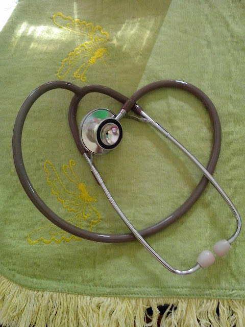
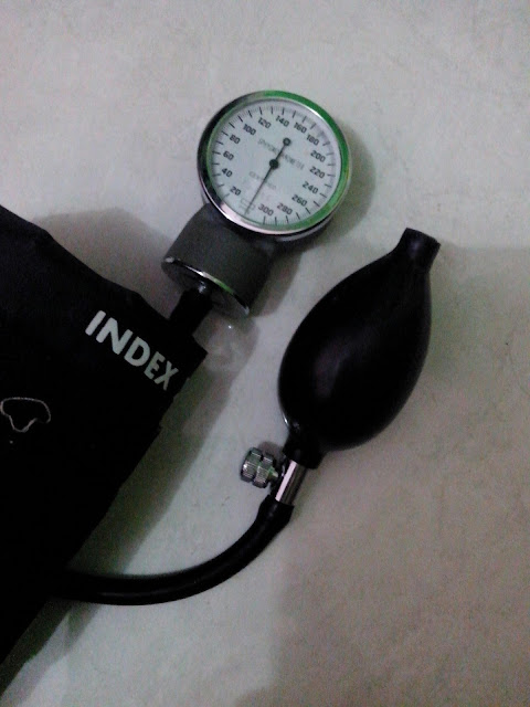
• How Stethoscope Works....

    A stethoscope is a medical device for listening to sounds inside the body. The initial stethoscope was invented in the early 19th century by French physician Ren� Laennec, but was actually trying to achieve a rather different end: doctor-patient distance....

  • Care Plan For Decreased Cardiac Output...

    Nursing diagnosis for decreased cardiac output may be related to altered myocardial contractility, inotropic changes; alterations in rate, rhythm, electrical conduction; or structural changes, such as valvular defects and ventricular aneurysm. ...

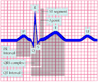
  • ECG Waveforms And Components

    The electrocardiogram (ECG) is a graphic recording ofelectric potentials generated by the heart.The signals are detected by means of metal electrodes attached to the extremities and chest wall and are then amplified and recorded by the electrocardiograph. ECG leads actually display the instantaneous differences in potential between these electrodes. ...

Showing posts with label fundamental. Show all posts

Thursday, December 19, 2013

Transmission of Hepatitis C

Hepatitis C is a disease with a significant global impact. According to the World Health Organization there are 170 million people infected with hepatitis C virus (HCV). There are considerable regional differences. In Europe and the United States chronic hepatitis C is the most common chronic liver disease. The majority of liver transplants performed in these regions are for chronic HCV. It is difficult to determine the number of new HCV infections, as most acute cases are not noticed clinically.
Parenteral exposure to the hepatitis C virus is the most efficient means of transmission. The majority of patients infected with HCV in Europe and the United States acquired the disease through intravenous drug use or blood transfusion, which has become rare since routine testing of the blood supply for HCV began. The following possible routes of infection have been identified in blood donors (in descending order of transmission risk):
  • Injection drug use
  • Blood transfusion
  • Sex with an intravenous drug user
  • Having been in jail more than three days
  • Religious scarification
  • Having been struck or cut with a bloody object
  • Pierced ears or body parts
  • Immunoglobulin injection
Very often in patients with newly diagnosed HCV infection no clear risk factor can be identified.
Factors that may increase the risk of HCV infection include greater numbers of sex partners, history of sexually transmitted diseases, and failure to use a condom. Whether underlying HIV infection increases the risk of heterosexual HCV transmission to an uninfected partner is unclear. The seroprevalence of HCV in MSM (men who have sex with men) ranges from about 4 to 8%, which is higher than the HCV prevalence reported for general European populations.
The risk of perinatal transmission of HCV in HCV RNA positive mothers is estimated to be 5% or less (Ohto 1994). Caesarean section has not been shown to reduce transmission. There is no evidence that breastfeeding is a risk factor.
Hemodialysis risk factors include blood transfusions, the duration of hemodialysis, the prevalence of HCV infection in the dialysis unit, and the type of dialysis. The risk is higher with in-hospital hemodialysis vs peritoneal dialysis.
Contaminated medical equipment, traditional medicine rites, tattooing, and body piercing are considered rare transmission routes.
There is some risk of HCV transmission for health care workers after unintentional needle-stick injury or exposure to other sharp objects.
Read More

Thursday, November 28, 2013

Overview of Respiratory Function

The major function of lungs and pulmonary circulation as the pulmonary system is to deliver oxygen (O2) to cells and remove carbon dioxide (CO2) from the cells (gas exchange). The adequacy of oxygenation and ventilation is measured by partial pressure of arterial oxygen (PaO2) and partial pressure of arterial carbon dioxide (PaCO2). The pulmonary system also functions as a blood reservoir for the left ventricle when it is needed to boost cardiac output; as a protector for the systemic circulation by filtering debris/particles; as a fluid regulator so water can be kept away from alveoli; and as a provider of metabolic functions such as surfactant production and endocrine functions.
Terminology in Respiratory
  • Alveolus—air sac where gas exchange takes place
  • Apex—top portion of the upper lobes of lungs
  • Base—bottom portion of lower lobes of lungs, located just above the diaphragm
  • Bronchoconstriction—constriction of smooth muscle surrounding bronchioles
  • Bronchus—large airways; lung divides into right and left bronchi
  • Carina—location of division of the right and left main stem bronchi
  • Cilia—hairlike projections on the tracheobronchial epithelium, which aid in the movement of secretions and removal of debris
  • Compliance—ability of the lungs to distend and change in volume relative to an applied change in pressure (eg, emphysema—lungs very compliant; fibrosis—lungs noncompliant or stiff)
  • Dead space—ventilation that does not participate in gas exchange; also known as wasted ventilation when there is adequate ventilation but no perfusion, as in pulmonary embolus or pulmonary vascular bed occlusion. Normal dead space is 150 mL.
  • Diaphragm—primary muscle used for respiration; located just below the lung bases, it separates the chest and abdominal cavities
  • Diffusion (of gas)—movement of gas from area of higher to lower concentration
  • Dyspnea—subjective sensation of breathlessness associated with discomfort, often caused by a dissociation between motor command and mechanical response of the respiratory system as in:
    • Respiratory muscle abnormalities (hyperinflation and airflow limitation from chronic obstructive pulmonary disease [COPD]).
    • Abnormal ventilatory impedance (narrowing airways and respiratory impedance from COPD or asthma).
    • Abnormal breathing patterns (severe exercise, pulmonary congestion or edema, recurrent pulmonary emboli).
    • Arterial blood gas (ABG) abnormalities (hypoxemia, hypercarbia).
  • Hemoptysis—coughing up of blood
  • Hypoxemia—PaO2 less than normal, which may or may not cause symptoms (Normal PaO2 is 80 to 100 mm Hg on room air.)
  • Hypoxia—insufficient oxygenation at the cellular level due to an imbalance in oxygen delivery and oxygen consumption (Usually causes symptoms reflecting decreased oxygen reaching the brain and heart.)
  • Mediastinum—compartment between lungs containing lymph and vascular tissue that separates left from right lung
  • Orthopnea—shortness of breath when in reclining position
  • Paroxysmal nocturnal dyspnea—sudden shortness of breath associated with sleeping in recumbent position
  • Perfusion—blood flow, carrying oxygen and CO2 that passes by alveoli
  • Pleura—serous membrane enclosing the lung; comprised of visceral pleura, covering all lung surfaces, and parietal pleura, covering chest wall and mediastinal structures, between which exists a potential space
  • Pulmonary circulation—network of vessels that supply oxygenated blood to and remove CO2-laden blood from the lungs
  • Respiration—inhalation and exhalation; at the cellular level, a process involving uptake of oxygen and removal of CO2 and other products of oxidation
  • Shunt—adequate perfusion without ventilation, with deoxygenated blood conducted into the systemic circulation, as in pulmonary edema, atelectasis, pneumonia, COPD
  • Surfactant—fluid secreted by alveolar cells that reduces surface tension of pulmonary fluids and aids in elasticity of pulmonary tissue
  • Ventilation—movement of air (gases) in and out of the lungs
  • Ventilation-perfusion (V/Q) imbalance or mismatch—imbalance of ventilation and perfusion; a cause for hypoxemia. V/Q mismatch can be due to:
    • Blood perfusing an area of the lung where ventilation is reduced or absent.
    • Ventilation of parts of lung that are not perfused.
Read More

Saturday, August 24, 2013

Basic Procedures That Must Be Understood By Every Nurse

Patients come to the hospital and other health facilities because they require skilled clinical observation and treatment. Millions of people hospitalized each year, and for the most part, it was a trying experience. Inpatient care dealing with patients' needs for privacy and control of his life. He should release at least part of the normal routine. He had to rely on you and your co-workers to meet basic needs. Depending on the complexity of health problems, he and his family may also require teaching, counseling, coordination of care, development of community support systems, and help in coping with changes related to health in his life.
Some broader aims of your care are helping the patient cope with restricted mobility; giving him a comfortable, stimulating environment; making sure his stay is free from hazards; promoting an uneventful recovery; and helping him return to his normal life.
Each time the patient's condition deter or prevent mobility, then your nursing goals include promoting independence by motivating him, helped him set goals, to prevent injury and complications of immobility, he teaches the skills needed, and encourage a positive body image, especially if he faces a long term or permanent immobility.
Besides weakening the patient, illness and any accompanying treatment may impair his judgment and contribute to accidents. Be alert to hazards in the patient's environment, and teach him and his family to recognize and correct them. When caring for a patient with restricted mobility, you must help him as he's moved, lifted, and transported. By using proper body mechanics and appropriate assistive devices, you can prevent injury, fatigue, and discomfort for the patient and yourself. To prevent complications, be sure to use correct positioning, meticulous skin care, assistive devices, and regular turning and range-of-motion exercises.
The first step toward rehabilitation typically is progressive ambulation, which should begin as soon as possible if necessary, using such assistive devices as a cane, crutches, or a walker. Demonstrating a technique such as transferring from a bed to a wheelchair during hospitalization helps the patient and his family to understand it. Allowing them to practice it under your supervision gives them the confidence to perform it at home. Encourage them to provide positive reinforcement to motivate the patient to work toward his goals.
 
Read More
Powered by Blogger.

Search This Blog