• How Stethoscope Works....

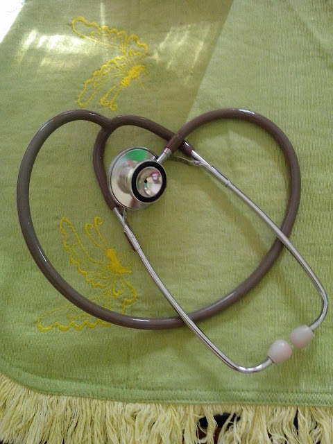
    A stethoscope is a medical device for listening to sounds inside the body. The initial stethoscope was invented in the early 19th century by French physician Ren� Laennec, but was actually trying to achieve a rather different end: doctor-patient distance....

  • Care Plan For Decreased Cardiac Output...

    Nursing diagnosis for decreased cardiac output may be related to altered myocardial contractility, inotropic changes; alterations in rate, rhythm, electrical conduction; or structural changes, such as valvular defects and ventricular aneurysm. ...

  • ECG Waveforms And Components

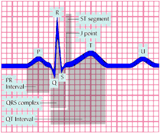
    The electrocardiogram (ECG) is a graphic recording ofelectric potentials generated by the heart.The signals are detected by means of metal electrodes attached to the extremities and chest wall and are then amplified and recorded by the electrocardiograph. ECG leads actually display the instantaneous differences in potential between these electrodes. ...

Friday, July 18, 2014

Achieving Healthy Mother, Baby, and Family Unit

INTRODUCTION TO MATERNITY NURSING
Providing care to childbearing families is aimed at the ideal of having every pregnancy result in a healthy mother, baby, and family unit. The nurse today faces many evolving and challenging issues in achieving this goal. Such advances as in vitro fertilization and embryo freezing have afforded people opportunities once thought impossible. An increasing number of high-risk pregnancies result from such factors as drug abuse, acquired immunodeficiency syndrome, late or no prenatal care, teenage pregnancies, and pregnancies in women older than age 35. Technologic advances in high-risk obstetric units, fetal monitoring, sonography, and neonatal intensive care units are now providing the means to improve maternal health and save fetuses and infants who would not have survived years ago.
Today's childbearing families have many options. The planned birth may take place in the traditional hospital setting, a birthing center, or at home. The primary care provider may be a physician, a certified nurse-midwife, or a lay midwife. Birth-related choices commonly include the use of labor, delivery, and recovery rooms or labor, delivery, recovery, and postpartum rooms; various birthing positions and analgesic methods; alternative pain-relief strategies such as hydrotherapy; and the decision to allow children and others to be present during labor and delivery. Regionalization of obstetric services has provided childbearing families with access to the technologic advances and skilled personnel capable of managing pregnancy or neonatal complications.
Economic changes in the health care climate have dramatically affected the practice of nursing as cost-containment considerations have shortened the hospital length of stay. Many hospitals have adopted a practice of 12- to 24-hour discharge after delivery coordinated with home health care follow-up.
This combination of advancing technology, pregnancy risk factors, and changing economics challenges the nurse to be a highly skilled clinician and outstanding communicator.
 
TERMINOLOGY USED IN MATERNITY NURSING
  • Gestation—pregnancy or maternal condition of having a developing fetus in the body.
  • Embryo—human conceptus up to the 10th week of gestation (8th week postconception).
  • Fetus—human conceptus from 10th week of gestation (8th week postconception) until delivery.
  • Viability—capability of living, usually accepted as 24 weeks, although survival is rare.
  • Gravida (G)—woman who is or has been pregnant, regardless of pregnancy outcome.
  • Nulligravida—woman who is not now and never has been pregnant.
  • Primigravida—woman pregnant for the first time.
  • Multigravida—woman who has been pregnant more than once.
  • Para (P)—refers to past pregnancies that have reached viability.
  • Nullipara—woman who has never completed a pregnancy to the period of viability. The woman may or may not have experienced an abortion.
  • Primipara—woman who has completed one pregnancy to the period of viability regardless of the number of infants delivered and regardless of the infant being live or stillborn.
  • Multipara—woman who has completed two or more pregnancies to the stage of viability.
  • Living children—refers to the number of children a woman has delivered who are living.
A woman who is pregnant for the first time is a primigravida and is described as Gravida 1 Para 0 (or G1P0). A woman who delivered one fetus carried to the period of viability and who is pregnant again is described as Gravida 2, Para 1. A woman with two pregnancies ending in abortions and no viable children is Gravida 2, Para 0.
Read More

Wednesday, July 16, 2014

STRENGTHENING PELVIC FLOOR MUSCLES

Stress incontinence, the most common kind of urinary incontinence in women, usually results from weakening of the urethral sphincter. In men, it may sometimes occur after a radical prostatectomy.
You can help male and female patients prevent or minimize stress incontinence by teaching pelvic floor (Kegel) exercises to strengthen the pubococcygeal muscles. Here's how.
Learning Kegel exercises
First, explain how to locate the muscles of the pelvic floor. Instruct the patient to tense the muscles around the anus, as if to retain stools.
To identify this area initially, teach the patient to tighten the muscles of the pelvic floor to stop the flow of urine while urinating and then to release the muscles to restart the flow. Once learned, these exercises can be done anywhere. Although Kegel exercises shouldn't be done while urinating, they can be done at any other time.

, ,
Establishing a regimen
Explain to the patient that contraction and relaxation exercises are essential to muscle retraining. Suggest that the patient start out by contracting the pelvic floor muscles for 5 seconds, relax for 5 seconds, and then repeat the procedure as often as needed.
Typically, the patient starts with 10 contractions in the morning and 10 at night, gradually increasing the relaxation and contraction time.
Advise the patient not to use stomach, leg, or buttock muscles. Also discourage leg crossing or breath holding during these exercises.
Read More

Sunday, June 22, 2014

Selection of Phlebotomy Venipuncture Site

Antecubital vein location varies slightly from person to person; however, two basic vein distribution arrangements referred to as the “H-shaped” and the “M-shaped” patterns are seen most often. The “H-shaped” pattern is so named because the most prominent veins in this pattern- the cephalic, cephalic median, median basilic, and basilic veins- are distributed on the arm in a way that resembles a slanted H. The most prominent veins of the M pattern- the cephalic, median cephalic, median basilic, and basilic veins- resemble the shape of an M. The H-shaped pattern is seen in approximately 70% of the population.
Factors in Vein Selection: Select the vein carefully. The brachial artery and several major nerves pass through the antecubital area. Accidental artery puncture and nerve injury are risks of venipuncture. Prioritizing veins can minimize the potential for accidental arterial puncture and nerve involvement. Typically, a tourniquet is used to aid in the selection of a vein unless specific tests require that a tourniquet not be used. A tourniquet is not necessary if veins are large and easily palpated. However, if only the basilic vein is visible without a tourniquet, one must be applied so the availability of safer veins (e.g. median and/or cephalic) can be assessed. Palpation is usually performed using the index finger. The collector’s thumb should not be used to palpate because it has a pulse beat. In addition to locating veins, the palpation pressure helps to differentiate veins from arteries, which pulsate, are more elastic, and have a thick wall.
Accidental Arterial Puncture: If during the procedure accidental arterial puncture is suspected (e.g. rapidly forming hematoma, rapid filling tube, and bright red blood), discontinue the venipuncture immediately. Remove the needle and apply direct forceful pressure to the puncture site for a minimum of 5 minutes until active bleeding has ceased. The nursing staff and physician must be notified and the incident documented according to institutional policy.
Consult with supervisory personnel to determine the suitability of the suspected arterial specimen for testing. If the specimen is acceptable it must be annotated that the specimen was an arterial specimen. In some cases different normal reference intervals are assigned to arterial blood. This information must be conveyed to the caregiver through Meditech Specimen Collection comment and Test Result comments.
Nerve Injury: If the patient feels a shooting, electric-like pain, or tingling or numbness proximal or distal to the venipuncture site, terminate the venipuncture and remove the needle immediately. Repeat the venipuncture in another site with a new sterile needle if needed. Document the incident and direct the patient to medical evaluation if indicated.
Read More

Friday, June 20, 2014

Take a look at the build up to the game from the inside

More Info

     
Emi's Stethoscope,
Here's what's trending on Twitter this week.
 

80.000 flags! Supporters at the Camp Nou tonight will find a Barça flag on their seats bit.ly/1he5agB pic.twitter.com/fxAjGVhqYJ
01 Apr
Exculsive images of the team's final training session on Monday and Dani Alves and Tata Martino's press conferencel
1
Related tweet
Samsung GS5 Punya Fitur Rahasia, Apa itu? goo.gl/xOYvCd
01 Apr
Kalau droiders sedang di pasar mencari smartphone baru, Samsung Galaxy S5 adalah salah satu yang jadi pilihan popular
Produits officiels de la ligue des champions de l'UEFA DISPONIBLES MAINTENANT sur votre boutique en ligne ! store.uefa.com/stores/uefa/
31 Mar
Shop for the latest football shirts, football boots, kits, fashion, Equipment & souvenirs on the UEFA Champions League Official Online Store
@einyasha We're glad that you've enjoyed XP! In case you haven't heard, here is some information about about XP: msft.it/6014gASp.
31 Mar
Get end of support information for Windows XP and find out what you need to know to upgrade to Windows 8.1 or find a new PC.
Amazing fans !! Thanks for the support tonight pic.twitter.com/ILejGDAnsy
01 Apr
1
Retweet
We're expecting a great atmosphere at OT tonight - these @FCBayernEN fans have been warming up en route. #MUFCBAYERN pic.twitter.com/aYQ9hGC1qR
01 Apr
1
Retweet
The Camp Nou 30 minutes before kick off #FCBlive #FCBAtleti pic.twitter.com/PbAifg48Ok
01 Apr
1
Retweet
Not working tonight but couldn't miss this one. pic.twitter.com/f2Zb81llFR
01 Apr
1
Retweet
Don't miss out. Stay up to date on what's happening.
Go to Twitter
Read More

Correcting Urinary Incontinence With Bladder Retraining

The incontinent patient typically feels frustrated, embarrassed, and hopeless. Fortunately, his problem can usually be corrected by bladder retraining—a program that aims to establish a regular voiding pattern. Follow these guidelines.
Assess elimination patterns
First, assess the patient's intake and voiding patterns and reason for each accidental voiding (such as a coughing spell). Use an incontinence monitoring record.
Establish a voiding schedule
Encourage the patient to void regularly, for example, every 2 hours. When he can stay dry for 2 hours, increase the interval by 30 minutes every day until he achieves a 3- to 4-hour voiding schedule. Teach the patient to practice relaxation techniques such as deep breathing, which help decrease the sense of urgency.
Record results and remain positive
Keep a record of continence and incontinence for about 5 days to help reinforce the patient's efforts to remain continent. Remember, both your own and your patient's positive attitudes are crucial to his successful bladder retraining.
Take steps for success
Here are some additional tips to boost the patient's success:
  • Be sure to locate the patient's bed near a bathroom or portable toilet. Leave a light on at night. If the patient needs assistance getting out of bed or a chair, promptly answer the call for help.
  • Teach the patient measures to prevent urinary tract infections, such as adequate fluid intake (at least 2,000 ml/day unless contraindicated), drinking cranberry juice to help acidify urine, wearing cotton underpants, and bathing with nonirritating soaps. If the patient has urge incontinence, cranberry juice is contraindicated.
  • Encourage the patient to empty his bladder completely before and after meals and at bedtime.
  • Advise him to urinate whenever the urge arises and never to ignore it.
  • Instruct the patient to take prescribed diuretics upon rising in the morning.
  • Advise him to limit the use of sleeping aids, sedatives, and alcohol; they decrease the urge to urinate and can increase incontinence, especially at night.
  • If the patient is overweight, encourage weight loss.
  • Suggest exercises to strengthen pelvic muscles.
  • Instruct the patient to increase dietary fiber to decrease constipation and incontinence.
  • Monitor the patient for signs of anxiety and depression.
  • Reassure the patient that periodic incontinent episodes don't mean that the program has failed. Encourage persistence, tolerance, and a positive attitude.
Read More

Wednesday, June 18, 2014

Phlebotomy Venipuncture Procedure : Guidelines to Prioritizing Draws

Specimen Priority:
“The urgency that the healthcare provider wants the specimen collected/run determines that order’s priority. We have three specimen priorities:
A. STAT (S) – this priority means that the order is to be collected and run right away, without delay. This status is used most often in emergency situations and should be the first specimens collected.
B. URG (U) – this is the urgent specimen priority and it is to be collected as soon as possible but not before a STAT. Urgent specimens are the middle priority, meaning they are important to be done quickly but not absolutely critical (i.e. STAT)
C. ROUTINE (R) – this is the lowest specimen priority. Routine specimens should be collected when you can but not before a STAT or URG.
- The last thing to understand about specimen priority is the concept of Timed Orders. Any one of the above three priorities may be ordered for a specific time of the day to be collected (i.e. URG for 2000 or ROUTINE for 2359). These are called TIMED ORDERS. When a specimen is requested for a specific time then the priority is superceded and the specimen should be collected at the requested time. All STAT and URG specimens (regardless of what time they are ordered for) go into their respective Collection Category (STAT or URG, see above). If a specimen is ordered as a ROUTINE priority for a specific time, it will go into that specific Collection Category time (i.e. 0830 or 1600). If a
specimen is ROUTINE priority and DOES NOT have a specific time requested it will go into the POOL Collection Category. Any specimens in the POOL Collection Category are of the lowest urgency, to be collected whenever you have time.”
NOTE: STATS and TIMED ORDERS are to be collected within a 15 minute window (15 minutes before
or after the ordered time). It is encouraged that these specimens are received in the laboratory within 15
minutes of being drawn.

 
Read More

Wednesday, June 11, 2014

Phlebotomy Venipuncture Procedure : Assembly of Supplies/ Safety

All needles and syringes must be taken out of the original package in the patient’s presence. Never
preassemble the vacutainer apparatus with a needle prior to use.
1. Vacutainer Eclipse
a. Holding both colored shields, twist and remove white shield (end to be screwed into holder).
b. Screw in holder.
c. Rotate safety shield back out of way.
2. Syringe Draw
a. Open Hypodermic Needle package and remove device.
b. Attach appropriate Hypodermic Needle to syringe by twisting the needle onto the syringe.
c. When transferring the specimen to a vacutainer tube, no associate is to handle a tube at any time
with their hand while inserting the transfer needle into the vacutainer. This includes stabilizing a
tube with one’s hand, in the phlebotomy tray, while inserting the needle. The hand that is not
holding the syringe is not to aid in specimen transfer. The associate may only use their hand to
remove the vacutainer tube from the transfer needle, while pulling the vacutainer tube away from
the transfer needle and either immediately placing the needle in another tube located on the
phlebotomy tray, or immediately activating the safety shield for discard, if the specimen transfer is
complete. If the specimen cannot be successfully transferred without the need for an associate to
use their “free” hand to hold or stabilize a vacutainer tube, then a transfer hub is to be utilized.
3. Butterfly “Push Button” with a SYRINGE
a. Open Butterfly Push Button package and remove device.
b. Remove luer cap and attach syringe
3
c. Remove the clear plastic needle sheath.
4. Butterfly “Push Button” with a VACUTAINER
a. Screw multiple adaptor into vacutainer.
b. Open Butterfly Push Button package and remove device.
c. Remove colored end of multiple sample adaptor and remove luer cap to attach butterfly.
d. Remove the clear plastic needle sheath.
Read More
Powered by Blogger.

Search This Blog