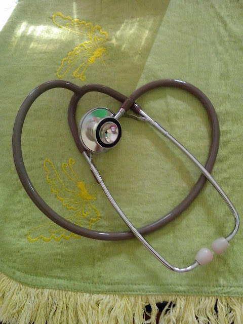
A stethoscope is a medical device for listening to sounds inside the body. The initial stethoscope was invented in the early 19th century by French physician Ren� Laennec, but was actually trying to achieve a rather different end: doctor-patient distance....
Wednesday, July 8, 2015
Cranial Nerve - Brainstem Function
Cranial nerve assessment is basically an assessment of brainstem function because nuclei of 10 of the 12 cranial nerves are located in the brainstem. The proximity of these nuclei to the reticular activating system (arousal center) located in the midbrain is the anatomic rationale for assessing cranial nerves in conjunction with LOC. Important neurological functions and protective reflexes are mediated by the cranial nerves and many functions are dependent on more than one nerve. Some of the cranial nerves have both motor and sensory functions.
The two cranial nerves that do not arise in the brainstem are the olfactory nerve (CN I) and the optic nerve (CN II). CN I is located in the medial frontal lobe and is responsible for the sense of smell. This can be difficult to assess in the younger child, so is often omitted unless there is specific concern that there has been damage in that area. Taste may also be affected with injuries to CN I. CN II is assessed by determining a child’s visual acuity. This may be done more formally with visual screening or more generally by noting if the child’s vision appears normal in routine activities.
Pupil size and response to direct light are mediated by CN II and the oculomotor nerve (CN III) as well as the sympathetic nervous system. Many things can affect the pupillary response in a child, including damage to the eye or the cranial nerves, pressure on the upper brainstem, local and systemic effects of certain drugs, anoxia, and seizures. Pupillary size varies with age and is determined by the amount of sympathetic input, which dilates the pupil and is balanced by the parasympathetic input on CN III, which constricts the pupil. Pupillary response in the eye that is being checked with direct light as well as the other pupil (consensual response) are significant in that they can point to where damage to nerves exists and are an objective clinical sign that can be followed over time .
 |
| Diagram of the base of the brain showing entrance and exits of the cranial nerves |
The two cranial nerves that do not arise in the brainstem are the olfactory nerve (CN I) and the optic nerve (CN II). CN I is located in the medial frontal lobe and is responsible for the sense of smell. This can be difficult to assess in the younger child, so is often omitted unless there is specific concern that there has been damage in that area. Taste may also be affected with injuries to CN I. CN II is assessed by determining a child’s visual acuity. This may be done more formally with visual screening or more generally by noting if the child’s vision appears normal in routine activities.
Pupil size and response to direct light are mediated by CN II and the oculomotor nerve (CN III) as well as the sympathetic nervous system. Many things can affect the pupillary response in a child, including damage to the eye or the cranial nerves, pressure on the upper brainstem, local and systemic effects of certain drugs, anoxia, and seizures. Pupillary size varies with age and is determined by the amount of sympathetic input, which dilates the pupil and is balanced by the parasympathetic input on CN III, which constricts the pupil. Pupillary response in the eye that is being checked with direct light as well as the other pupil (consensual response) are significant in that they can point to where damage to nerves exists and are an objective clinical sign that can be followed over time .
Modified Glasgow Coma Scale For Infants And Children
Modified Glasgow Coma Scale for infants and children. Coma scoring system appropriate for pediatric patients.
Obtained from Marcoux (2005) [24]
Obtained from Marcoux (2005) [24]
Activity
|
Score
|
Infant/non-verbal child (<2 years)
|
Verbal child/adult (>2 years)
|
|
|---|---|---|---|---|
Eye Opening |
4 |
Spontaneous |
Spontaneous |
|
3 |
To Speech |
To verbal stimuli |
||
2 |
To Pain Only |
To Pain Only |
||
1 |
No Response |
No Response |
||
Motor Response |
6 |
Normal/ spontaneous movement |
Obeys commands |
|
5 |
Withdraws to touch |
Localizes pain |
||
4 |
Withdraws to pain |
Flexion withdrawal |
||
3 |
Abnormal flexion (decorticate) |
Abnormal flexion |
||
2 |
Extension (decerebrate) |
Extension (decerebrate) |
||
1 |
No response |
No response |
||
2–5 years
|
> 5 years
|
|||
Verbal Response |
5 |
Cries appropriately, coos |
Appropriate words |
Oriented |
4 |
Irritable crying |
Inappropriate words |
Confused |
|
3 |
Inappropriate screaming / crying |
Screams |
Inappropriate |
|
2 |
Grunts |
Grunts |
Incomprehensible |
|
1 |
No Response |
No Response |
No Response | |
Sunday, July 5, 2015
Developmental Screening Tools Commonly Used To Assess Child Development
Posted by
Channel Maymoon
Labels:
Adolescent,
Child,
Infant,
Neonate,
Neurological Assessment
at
7:59 PM
Developmental screening tools commonly used to assess child development. Data from references: Behrman et al. (2004) [4] and Wong et al. (2000) [35]
Tool name
|
Revised Denver
developmental
screening test
(Denver II)
|
Prescreening
developmental questionnaire
R-PDQ)
|
Developmental
profile II
|
Draw a person
(DAP) test
|
|---|---|---|---|---|
Author |
Frankenburg [13] |
Frankenburg et al. [14] |
Alpern et al. [1] |
Goodenough [15] |
Items scored |
Gross motor Fine motor Language Personal-social |
Parent answered prescreen of items on Denver II |
Physical Self-help Social Academic Communication |
Score for body parts |
Age range |
Birth–6 years |
Birth–6 years |
Birth–7 years |
5–17 years |
Interview |
Parent/child |
Parent only |
Parent/child |
Child only |
Testing time |
30–40 minutes |
15–20 min |
20–40 min |
As needed |
Training/certified |
Yes |
Self-instruction |
Self-instruction |
Self-instruction |
Pros/cons |
Range of items Identify child’s strengths/weakness Validity tested Cultural bias Teaching tool |
Parent report Can rescreen If delays administer Denver II |
Range of items Low rate of sensitivity |
Nonverbal Nonthreatening Cultural unbias Few items to score Gives IQ score |
Friday, July 3, 2015
Age-appropriate Neuroassessment
Age-appropriate neuroassessment table. A brief guide to developmental milestones in children from infancy to age 12 years as a guide when performing a neurological assessment (Phoenix Children’s Hospital)
Read More
Age
|
Gross Motor
|
Fine Motor
|
Personal/social
|
Language
|
|---|---|---|---|---|
Newborn
|
Head down with ventral suspension
Flexion Posture
Knees under abdomen-pelvis high
Head lag complete
Head to one side prone
|
Hands closed
Cortical Thumbing (CT)
|
With sounds, quiets if
crying; cries if quiet;
startles; blinks
|
Crying only
monotone
|
4 weeks
|
Lifts chin briefly (prone)
Rounded back sitting
head up momentarily
Almost complete head lag
|
Hands closed (CT)
|
Indefinite stare
at surroundings
Briefly regards toy
only if brought
in front of eyes
and follows only
to midline
Bell sound
decreases activity
|
Small, throaty noises
|
6 weeks
|
In ventral suspension head up
momentarily in same plane as body
Prone: pelvis high but knees no
longer under abdomen
|
Hands open 25%
of time
|
Smiles
|
Social smile
(1st cortical input)
|
2 months
|
Ventral suspension; head in same
plane as body
Lifts head 45° (prone) on flexed
forearms
Sitting, back less rounded, head
bobs forward
Energetic arm movements
|
Hands open most of
the time (75%)
Active grasp of toy
|
Alert expression
Smiles back
Vocalizes when talked to
Follows dangled toy
beyond midline
Follows moving person
|
Cooing
Single vowel sounds
(ah. eh, uh)
|
3 months
|
Ventral suspension; head in same
plane as body
Lifts head 45° (prone) on flexed
forearms
Sitting, back less rounded, head
bobs forward
Energetic arm movements
|
Hands open most of
the time (75%)
Active grasp of toy
|
Smiles spontaneously
Hand regard
Follows dangled toy 180°
Promptly looks at object
in midline
Glances at toy put in hand
|
Chuckles
“Talk back” if
examiner nods head
and talks
Vocalizes with two
different syllables
(a-a. oo-oo)
|
4 months
|
Head to 90° on extended forearms
Only slightly head lag at beginning
of movement
Bears weight some of time on
extended legs if held standing
Rolls prone to supine
Downward parachute
|
Active play with
rattles
Crude extended reach
and grasp
Hands together
Plays with fingers
Toys to mouth when
supine
|
Body activity increased
at sight of toy
Recognizes bottle and
opens mouth
For nipple (anticipates
feeding with excitement)
|
Laughs out loud
increasing
inflection
No tongue thrust
|
6 months
|
Bears full weight on legs if held
standing
Sits alone with minimal support
Pivots in prone
Rolls easily both ways
Anterior proppers
|
Reaches for toy
Palmar grasp of cube
Lifts cup by handle
Plays with toes
|
Displeasure at removal
of toy
Puts toy in mouth if
sitting
|
Shy with
strangers
Imitates cough and
protrusion of tongue
Smiles at mirror
image
|
7 months
|
Bears weight on one hand prone
Held standing, bounces
Sit on hard surface leaning on
hands
|
|
Stretches arms to be taken
Keeps mouth closed if offered
more food than
wants
Smiles and pats at mirror
|
Murmurs “mom”
especially if
crying
Babbles easily
(M’s, D’s, B’s, L’s)
Lateralizes sound
|
9 months
|
Sits steadily for 15 min on hard surface
Reciprocally crawls
Forward parachute
|
Picks up small objects
with index finger
and thumb
(Pincer grasp)
|
Feeds cracker neatly
Drinks from cup with
help
|
Listens to conversation
Shouts for attention
Reacts to “strangers”
|
10 months
|
Pulls to stand
Sits erect and steadily (indefinitely)
Sitting to prone
Standing: collapses and creeps on
hands knees easily
Prone to sitting easily
Cruises – laterally
Squats and stoops – does not
recover to standing position
|
Pokes with index
finger, prefers small
to large objects
|
Nursery games
(i.e., pat-a-cake),
picks up dropped bottle,
waves bye-bye
|
Will play peek-a-boo
and pat-a-cake
to verbal command
Says Mama,
Dada appropriately,
finds the hidden toy
(onset visual
memory)
|
12 months
|
Sitting; pivots to pick up object
Walks, hands at shoulder height
Bears weight alone easily
momentarily
|
Easy pinch grasp with
arm off table
Independent release
(ex: cube into cup)
Shows preference for
one hand
|
Finds hidden toy under
cup
Cooperated with dressing
Drinks from cup with two
hands
Marks with crayon on
paper
Insists on feeding self
|
One other word
(noun) besides
Mama, Dada
(e.g., hi, bye, cookie)
|
13 months
|
Walks with one hand
|
Mouthing very little
Explores objects with
fingers
Unwraps small cube
Imitates pellet bottle
|
Helps with dressing
Offers toy to mirror image
Gives toy to examiner
Holds cup to drink, tilting
head
Affectionate
Points with index finger
Plays with washcloth,
bathing
Finger-feeds well, but
throws dishes on floor
Appetite decreases
|
Three words besides
Mama, Dada
Larger receptive
language than
expressive
|
14 months
|
Few steps without support
|
Deliberately picks up
two small blocks in
one hand
Peg out and in
Opens small square
box
|
Should be off bottle
Puts toy in container if
asked
Throws and plays ball
|
Three to four words
expressively
minimum
|
15 months
|
Creeps up stairs
Kneels without support
Gets to standing without support
Stoop and recover
Cannot stop on round corners suddenly
Collapses and catches self
|
Tower of two cubes
“Helps” turn pages
of book
Scribbles in imitation
Completes round peg
board with urging
|
Feeds self fully leaving
dishes on tray
Uses spoon turning upside
down, spills much
Tilts cup to drink, spilling
some
Helps pull clothes off
Pats at picture in book
|
Four to six words
Jargoning
Points consistently to
indicate wants
|
18 months
|
Runs stiffly
Rarely falls when walking
Walks upstairs (one hand held-one
step at a time)
Climbs easily
Walks, pulling toy or carrying doll
Throws ball without falling
Knee flexion seen in gait
|
Tower of three to four
cubes
Turns pages two to
three at a time
Scribbles
spontaneously
Completes round peg
board easily
|
Uses spoon without rotation
but still spills
May indicate wet pants
Mugs doll
Likes to take off shoes and
socks
Knows one body part
Very negative oppositions
|
One-step commands
10-15 words
Knows “hello” and
“thank you”
More complex
‘jargon’ rag
Attention span
1 min
Points to one picture
|
21 months
|
Runs well, falling some tires
Walks downstairs with one hand
held, one step at a time
Kicks large ball with demonstration
Squats in play
Walks upstairs alternating feet with
rail held
|
Tower of five to six
cubes
Opens and closes small
square box
Completes square peg
board
|
May briefly resist bathing
Pulls person to show something
Handles cup will Removes
some clothing purposefully
Asks for food and
drink Communicates toilet
needs helps wit h simple
household tasks 3 body
parts
|
Knows 15–20 words
and combines
2–3 words
Echoes 2 or more
Knows own name
Follows associate
commands
|
24 months
|
Rarely falls when running
Walks up and down stairs alone
one-step-at-a time
Kicks large ball without
demonstration
Claps hands
Overthrow hand
|
Tower of six to seven
cubes
Turns book pages
singly
Turns door knob
Unscrews lid
Replaces all cubes in
small box
Holds glass securely
with one hand
|
Uses spoon, spilling little
Dry at night
Puts on simple garment
Parallel play
Assists bathing
Likes to wash 6 dry hands
Plays with food
+ body parts
Tower of 8. Helps put
things away
|
Attention span 2 min
Jargon discarded
Sentences of two to
three words
Knows 50 words
Can follow two-step
commands (ain’t)
Refers to self by
name
Understands and
asks for “more”
Asks for food by
name
Inappropriately uses
personal pronouns
(e.g., me want)
Identifies three
pictures
|
3–5 years
|
Pedals tricycle
Walks up stairs alternating feet
Tip toe
Jump with both feet
|
Copies circles
Uses overhand throw
|
Group play
Can take turns
|
Uses three-word
sentences
|
5–12 years
|
Activities of daily living
|
Printing and cursive
writing
|
Group Sports
|
Reads and understands
content
Spells words
|
Wednesday, June 24, 2015
Surgical Risk Factors And Preventive Strategies : Perioperative Nursing
Obesity
Poor Nutrition
Fluid and Electrolyte Imbalance
Aging
Presence of Cardiovascular Disease
Therapeutic Approach
Presence of Pulmonary and Upper Respiratory Disease
Concurrent or Prior Pharmacotherapy
Read More
Danger
-
Increases the difficulty involved in the technical aspects of performing surgery; risk for wound dehiscence is greater
-
Increases the likelihood of infection because of compromised tissue perfusion
-
Increases the potential for postoperative pneumonia and other pulmonary complications because obese patients chronically hypoventilate
-
Increases demands on the heart, leading to cardiovascular compromise
-
Increases the risk for airway complications
-
Alters the response to many drugs and anesthetics
-
Decreases the likelihood of early ambulation
Therapeutic Approach
-
Encourage weight reduction if time permits.
-
Anticipate postoperative obesity-related complications.
-
Be extremely vigilant for respiratory complications.
-
Carefully splint abdominal incisions when moving or coughing.
-
Be aware that some drugs should be dosed according to ideal body weight versus actual weight (owing to fat content); otherwise, an overdose may occur (digoxin [Lanoxin], lidocaine [Xylocaine], aminoglycosides, and theophylline [Theo-Dur]).
-
Avoid intramuscular injections in morbidly obese individuals (I.V. or subcutaneous routes preferred).
-
Never attempt to move an impaired patient without assistance or without using proper body mechanics.
-
Obtain a dietary consultation early in the patient's postoperative course.
Danger
-
Greatly impairs wound healing (especially protein and calorie deficits and a negative nitrogen balance)
-
Increases the risk of infection
Therapeutic Approach
-
Any recent (within 4 to 6 weeks) weight loss of 10% of the patient's normal body weight or decreased serum albumin should alert the health care staff to poor nutritional status and the need to investigate as to the cause of the weight loss.
-
Attempt to improve nutritional status before and after surgery. Unless contraindicated, provide a diet high in proteins, calories, and vitamins (especially vitamins C and A); this may require enteral and parenteral feeding.
-
Review a serum prealbumin level to determine recent nutritional status.
-
Recommend repair of dental caries and proper mouth hygiene to prevent infection.
Fluid and Electrolyte Imbalance
Danger
Can have adverse effects in terms of general anesthesia and the
anticipated volume losses associated with surgery, causing shock and cardiac
dysrhythmias
Patients undergoing major abdominal operations
(such as colectomies and aortic repairs) often experience a massive fluid shift
into tissues around the operative site in the form of edema (as much as 1 L or
more may be lost from circulation). Watch for the fluid shift to reverse (from
tissue to circulation) around the third postoperative day. Patients with heart
disease may develop failure due to the excess fluid “load.”
Therapeutic Approach
-
Assess the patient's fluid and electrolyte status.
-
Rehydrate the patient parenterally and orally as prescribed.
-
Monitor for evidence of electrolyte imbalance, especially Na+, K+, Mg++, Ca++.
-
Be aware of expected drainage amounts and composition; report excess and abnormalities.
-
Monitor the patient's intake and output; be sure to include all body fluid losses.
Danger
-
Potential for injury is greater in older people
-
Be aware that the cumulative effect of medications is greater in the older patient
-
Medications in the usual dosages, such as morphine, may cause confusion, disorientation, and respiratory depression
Therapeutic Approach
-
Consider using lesser doses for desired effect.
-
Anticipate problems from chronic disorders such as anemia, obesity, diabetes, hypoproteinemia.
-
Adjust nutritional intake to conform to higher protein and vitamin needs.
-
When possible, cater to set patterns in older patients, such as sleeping and eating.
Danger
-
May compound the stress of anesthesia and the operative procedure
-
May result in impaired oxygenation, cardiac rhythm, cardiac output, and circulation
-
May also produce cardiac decompensation, sudden arrhythmia, thromboembolism, acute myocardial infarction (MI), or cardiac arrest
Therapeutic Approach
-
Frequently assess heart rate and blood pressure (BP) and hemodynamic status and cardiac rhythm if indicated.
-
Avoid fluid overload (oral, parenteral, blood products) because of possible MI, angina, heart failure, and pulmonary edema.
-
Prevent prolonged immobilization, which results in venous stasis. Monitor for potential deep vein thrombosis (DVT) or pulmonary embolus.
-
Encourage position changes but avoid sudden exertion.
-
Use antiembolism stockings and/or sequential compression device intraoperatively and postoperatively.
-
Note evidence of hypoxia and initiate therapy.
Presence of Diabetes Mellitus
Danger
-
Hypoglycemia may result from food and fluid restrictions and anesthesia.
-
Hyperglycemia and ketoacidosis may be potentiated by increased catecholamines and glucocorticoids due to surgical stress.
-
Chronic hyperglycemia results in poor wound healing and susceptibility to infection.
-
Recognize the signs and symptoms of ketoacidosis and hypoglycemia, which can threaten an otherwise uneventful surgical experience. Dehydration also threatens renal function.
-
Monitor blood glucose and be prepared to administer insulin, as directed, or treat hypoglycemia.
-
Confirm what medications the patient has taken and what has been held. Facility protocol and provider preference varies, but the goal is to prevent hypoglycemia. If the patient is NPO, oral agents are usually withheld and insulin may be ordered at 75% of the usual dose.
-
Reassure the diabetic patient that when the disease is controlled, the surgical risk is no greater than it is for the nondiabetic patient.
Presence of Alcoholism
Danger
The additional problem of malnutrition may be present in the
presurgical patient with alcoholism. The patient may also have an increased
tolerance to anesthetics.
Therapeutic Approach
-
Note that the risk of surgery is greater for the patient who has chronic alcoholism.
-
Anticipate the acute withdrawal syndrome within 72 hours of the last alcoholic drink.
Danger
Chronic pulmonary illness may contribute to hypoventilation,
leading to pneumonia and atelectasis. Surgery may be contraindicated in the
patient who has an upper respiratory infection because of the possible advance
of infection to pneumonia and sepsis.
Therapeutic Approach
-
Patients with chronic pulmonary problems, such as emphysema or bronchiectasis, should be evaluated and treated prior to surgery to optimize pulmonary function with bronchodilators, corticosteroids, and conscientious mouth care, along with a reduction in weight and smoking and methods to control secretions.
-
Opioids should be used cautiously to prevent hypoventilation. Patient-controlled analgesia is preferred.
-
Oxygen should be administered to prevent hypoxemia (low liter flow in chronic obstructive pulmonary disease).
Danger
Hazards exist when certain medications are given concomitantly with
others (eg, interaction of some drugs with anesthetics can lead to hypotension
and circulatory collapse). This also includes the use of many herbal substances.
Although herbs are natural products, they can interact with other medications
used in surgery.
Therapeutic Approach
-
An awareness of drug therapy is essential.
-
Notify the health care provider and anesthesiologist if the patient is taking any of the following drugs:
-
Certain antibiotics
-
Antidepressants, particularly monoamine oxidase inhibitors, and St. John's wort, an herbal product
-
Phenothiazines
-
Diuretics, particularly thiazides
-
Steroids
-
Anticoagulants, such as warfarin or heparin; or medications or herbals that may affect coagulation, such as aspirin, feverfew, ginkgo biloba, nonsteroidal anti-inflammatory drugs, ticlopidine (Ticlid), and clopidogrel (Plavix)
-
Friday, June 19, 2015
Diagnostic Imaging and Testing in Neurological Assessment - Pediatric Neurosurgery Patient
Posted by
Channel Maymoon
Labels:
Diagnostic Imaging and Testing,
Neurological Assessment,
Pediatric Neurosurgery
at
7:19 PM
Diagnostic imaging and other diagnostic tests play an important role in understanding the nature of neurological disorders. Advances in medicine, technology, and pharmacology have contributed to safer outcomes for children who may need sedation for diagnostic tests. Imaging or other tests may be performed to obtain a baseline for future studies.
In general, radiographic or digital imaging is looking at brain structure, while other diagnostic tests like electroencephalogram (EEG), single photon emission computed tomography scanning (SPECT),
nuclear medicine scans, and Wada tests are evaluating specific functions of the brain. Positron emission tomography (PET) scans look at metabolic function and utilization of glucose by the brain. Newer technologies allow for the evaluation of cerebral blood flow and brain perfusion. Some tests serve both diagnostic and therapeutic outcomes are:
nuclear medicine scans, and Wada tests are evaluating specific functions of the brain. Positron emission tomography (PET) scans look at metabolic function and utilization of glucose by the brain. Newer technologies allow for the evaluation of cerebral blood flow and brain perfusion. Some tests serve both diagnostic and therapeutic outcomes are:
- X-rays of the skull and vertebral column; X-rays to look at boney structures of the skull and spine, fractures, integrity of the spinal column, presence of calcium intra-cranially. Patient should be immobilized in a collar for transport if there is a question of spinal fracture.
- Cranial ultrasound; Doppler sound waves to image through soft tissue. In infants can only be used if fontanel is open. No sedation or intravenous access needed. Used to follow ventricle size/bleeding in neonates/infants.
- Computerized tomography with/without contrast; Differentiates tissues by density relative to water with computer averaging and mathematical reconstruction of absorption coefficient measurements. Non-invasive unless contrast is used or sedation needed. Complications include reaction to contrast material or extravasation at injection site.
- Computerized tomography - bone windows and/or threedimensional reconstruction; Same as above with software capabilities to subtract intracranial contents to look specifically at bone and reconstruct the skull or vertebral column in a three-dimensional model. No changes in study for patient. Used for complex skull and vertebral anomalies to guide surgical decision-making.
- Cerebral angiography; Intra-arterial injection of contrast medium to visualize blood vessels; transfemoral approach most common; occasionally brachial or direct carotid is used. Done under deep sedation or anesthesia; local reaction or hematoma may occur; systemic reactions to contrast or dysrhythmias; transient ischemia or vasospasm; patient needs to lie flat after and CMS checks of extremity where injection was done are required.
- MRI with or without contrast (gadolinium); Differentiates tissues by their response to radio frequency pulses in a magnetic field; used to visualize structures near bone, infarction, demyelination and cortical dysplasias. No radiation exposure; screened prior to study for indwelling metal, pacemakers, braces, electronic implants; sedation required for young children because of sounds and claustrophobia; contrast risks include allergic reaction and injection site extravasation.
- MRA MRV; Same technology as above used to study flow in vessels; radiofrequency signals emitted by moving protons can be manipulated to create the image of vascular contrast. In some cases can replace the need for cerebral angiography; new technologies are making this less invasive study more useful in children with vascular abnormalities.
- Functional MRI; Technique for imaging activity of the brain using rapid scanning to detect changes in oxygen consumption of the brain; changes can reflect increased activity in certain cells. Used in patients who are potential candidates for epilepsy surgery to determine areas of cortical abnormality and their relationship to important cortex responsible for motor and speech functions.
- SPECT; Nuclear medicine study utilizing injection of isotopes and imaging of brain to determine if there is increased activity in an area of abnormality; three-dimensional measurements of regional blood flow. Often used in epilepsy patients to diagnoses areas of cerebral uptake during a seizure (ictal SPECT) or between seizures (intraictal SPECT).
- SISCOM; Utilizing the technology of SPECT with MRI to look at areas of increased uptake in conjunction with MRI images of the cortex and cortical surface. No significant difference for patient; software as well as expertise of radiologist is used to evaluate study.
- PET; Nuclear medicine study that assesses perfusion and level of metabolic activity of both glucose and oxygen in the brain; radiopharmaceuticals are injected for the study. Patient should avoid chemicals that depress or stimulate the CNS and alter glucose metabolism (e.g., caffeine); patient may be asked to perform certain tasks during study.
- EEG Routine Ambulatory Video; Records gross electrical activity across surface of brain; ambulatory EEG used may be used for 24–48 h with data downloaded after study; video combines EEG recording with simultaneous videotaping. Success of study dependent on placement and stability of electrodes and ability to keep them on in children; routine studies often miss actual seizures but background activity can be useful information.
- Evoked responses;SSER;VER;BAER ; Measure electrical activity in specific sensorypathways in response to external stimuli; signal average produces waveforms that have anatomic correlates according to the latency of wave peaks. Results can vary depending on body size, age and characteristics of stimuli; sensation for each test will be different for patient – auditory clicks (BAER), strobe light (VER), or electrical current on skin – somatosensory (SSER).
Sunday, September 7, 2014
Maternal and Neonatal Care
Because of its profound emotional implications for mother and
child, maternal-neonatal care requires expertise that goes beyond clinical
skills. Such care must combine clinical competence, sensitivity, and good
judgment. It must consider the patient's sexuality and self-image and recognize
changing social attitudes and values—especially those concerning conventional
and alternative methods of conception and childbirth.
More than 4 million infants are born in the United States each
year. Many are born with considerably less medical intervention than was
customary in previous decades, and many were conceived with considerably more
intervention. As a result, nurses today must be prepared to implement or assist
with a wide range of procedures.
If you're working with a pregnant patient, you'll need to use your
teaching skills. For instance, you may be called on to organize and direct
natural childbirth classes or to teach the mother-to-be how to breathe and
control pain during childbirth. You may teach fathers and other support persons
to participate in childbirth by providing comfort and direction.
You may also be asked to give information about childbirth options.
Although most births still occur in a hospital, many parents inquire about
delivery in a birth center. Usually located on the maternity unit of a hospital
or sponsored by a childbirth association, a birth center combines the advantages
of a homelike setting with the emergency medical and nursing interventions
available in a hospital. Today's nurse may staff or direct the birth
center.
Historically, the midwife has been a fixture in remote or poor
communities. Today's professional nurse-midwife, however, brings advanced
technical skills and certification to diverse communities—urban center to
country town alike. She may work in collaboration with—or be supervised by—a
physician or a group. In some areas, she may even practice independently. In
fact, several states permit insurers to make direct payment to the nurse-midwife
for her services.
Accompanying the changes in maternity care are changes in neonatal
care—thanks to advanced knowledge and techniques for improving fetal
monitoring and promoting neonatal survival. New clinical evaluation methods,
combined with new electronic and biochemical monitoring techniques, allow
improved neonatal care. To make use of these advances, you must be familiar with
neonatal physiology, procedures, and equipment.
Subscribe to:
Posts (Atom)
Powered by Blogger.


