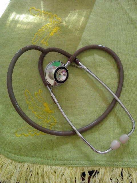
A stethoscope is a medical device for listening to sounds inside the body. The initial stethoscope was invented in the early 19th century by French physician Ren� Laennec, but was actually trying to achieve a rather different end: doctor-patient distance....
Thursday, December 19, 2013
Transmission of Hepatitis C
- Injection drug use
- Blood transfusion
- Sex with an intravenous drug user
- Having been in jail more than three days
- Religious scarification
- Having been struck or cut with a bloody object
- Pierced ears or body parts
- Immunoglobulin injection
Saturday, December 7, 2013
Simple music from John Lennon - Imagine (official video)
Imagine - John Lennon, A song about humanism.
Monday, December 2, 2013
Signs and Symptoms of Hydrocephalus
The signs and symptoms of hydrocephalus in infants and children vary depending on their age, the degree of hydrocephalus at presentation, the primary etiology, and the time over which the hydrocephalus develops. Ventriculomegaly can progress without obvious signs of increased intracranial pressure because of the plasticity of the infant brain and the ability of the cranium to expand.
In full-term infants, signs often include macrocephaly and progressively increasing occipital frontal head circumference, crossing percentile curves. Normal head circumference for a full-term infant is 33–36 cm at birth. A normal head circumference increases by approximately 2 cm/month during the first 3 months, by 1.5 cm/month during the 4th and 5th months, and by about 0.5 cm/month from months 6–12.
Signs and symptoms of hydrocephalus in children:
Premature infants
- Apnea
- Bradycardia
- Hypotonia
- Acidosis
- Seizures
- Rapid head growth
- Tense fontanel
- Splayed cranial sutures
- Vomiting
- Sunsetting eyes
Full-term infants
- Macrocephaly
- Rapid head growth
- Decreased feeding
- Increased drowsiness
- Tense fontanel
- Vomiting
- Distended scalp veins
- Splayed cranial sutures
- Poor head control
- Parinaud’s sign
- Sunsetting eyes
- Frontal bossing
Toddlers and older
- Headache
- Nausea
- Vomiting
- Irritability
- Lethargy
- Delayed development
- Decreased school performance
- Behavioral disturbance
- Papilledema
- Parinaud’s sign
- Sunsetting eyes
- Bradycardia
- Hypertension
- Irregular breathing patterns
Saturday, November 30, 2013
Nurse’s Ethical Duty in Wound Care
- First, the patient’s interests are placed above the personal interest of the nurse. If this duty is overlooked or forgotten, the contract (standard of practice) among the health-care provider, the health-care organization, and the patient is broken. — Example: The health-care provider conducts a seminar and needs wound photographs to supplement the written and verbal components of the presentation. The provider takes photographs of the patient’s wounds solely for the purpose of using them in the seminar. The only reason for taking these photographs is for the convenience of the health-care provider, and therefore the activity is actually for the nurse’s personal interest and not for the patient’s best interest. The patient would need to grant the nurse informed consent to use the photographs to avoid any consideration that the photographs are for personal interest. The nurse would need to assure the patient that any refusals on the patient’s part would have no effect on the nurse-patient relationship or the patient’s treatment.
- The patient’s privacy is protected from another individual’s or society’s desire to know details of the patient’s treatment. It is the health-care provider’s responsibility to have a complete understanding of the legal rights of all involved. It is ultimately the responsibility of the nurse to know the legal rights of the patient, family, and health-care provider. However, in many areas of the world, the general public does not have any legal right to knowledge concerning the patient’s care, progress, or prognosis. The health-care provider must identify if the health-care organization has a policy or procedure concerning this challenge.
- Does the health-care provider have a duty to treat the patient who has a wound(s)?
Thursday, November 28, 2013
Overview of Respiratory Function
- Alveolus—air sac where gas exchange takes place
- Apex—top portion of the upper lobes of lungs
- Base—bottom portion of lower lobes of lungs, located just above the diaphragm
- Bronchoconstriction—constriction of smooth muscle surrounding bronchioles
- Bronchus—large airways; lung divides into right and left bronchi
- Carina—location of division of the right and left main stem bronchi
- Cilia—hairlike projections on the tracheobronchial epithelium, which aid in the movement of secretions and removal of debris
- Compliance—ability of the lungs to distend and change in volume relative to an applied change in pressure (eg, emphysema—lungs very compliant; fibrosis—lungs noncompliant or stiff)
- Dead space—ventilation that does not participate in gas exchange; also known as wasted ventilation when there is adequate ventilation but no perfusion, as in pulmonary embolus or pulmonary vascular bed occlusion. Normal dead space is 150 mL.
- Diaphragm—primary muscle used for respiration; located just below the lung bases, it separates the chest and abdominal cavities
- Diffusion (of gas)—movement of gas from area of higher to lower concentration
- Dyspnea—subjective sensation of breathlessness associated with discomfort, often caused by a dissociation between motor command and mechanical response of the respiratory system as in:
- Respiratory muscle abnormalities (hyperinflation and airflow limitation from chronic obstructive pulmonary disease [COPD]).
- Abnormal ventilatory impedance (narrowing airways and respiratory impedance from COPD or asthma).
- Abnormal breathing patterns (severe exercise, pulmonary congestion or edema, recurrent pulmonary emboli).
- Arterial blood gas (ABG) abnormalities (hypoxemia, hypercarbia).
-
- Hemoptysis—coughing up of blood
- Hypoxemia—PaO2 less than normal, which may or may not cause symptoms (Normal PaO2 is 80 to 100 mm Hg on room air.)
- Hypoxia—insufficient oxygenation at the cellular level due to an imbalance in oxygen delivery and oxygen consumption (Usually causes symptoms reflecting decreased oxygen reaching the brain and heart.)
- Mediastinum—compartment between lungs containing lymph and vascular tissue that separates left from right lung
- Orthopnea—shortness of breath when in reclining position
- Paroxysmal nocturnal dyspnea—sudden shortness of breath associated with sleeping in recumbent position
- Perfusion—blood flow, carrying oxygen and CO2 that passes by alveoli
- Pleura—serous membrane enclosing the lung; comprised of visceral pleura, covering all lung surfaces, and parietal pleura, covering chest wall and mediastinal structures, between which exists a potential space
- Pulmonary circulation—network of vessels that supply oxygenated blood to and remove CO2-laden blood from the lungs
- Respiration—inhalation and exhalation; at the cellular level, a process involving uptake of oxygen and removal of CO2 and other products of oxidation
- Shunt—adequate perfusion without ventilation, with deoxygenated blood conducted into the systemic circulation, as in pulmonary edema, atelectasis, pneumonia, COPD
- Surfactant—fluid secreted by alveolar cells that reduces surface tension of pulmonary fluids and aids in elasticity of pulmonary tissue
- Ventilation—movement of air (gases) in and out of the lungs
- Ventilation-perfusion (V/Q) imbalance or mismatch—imbalance of ventilation and perfusion; a cause for hypoxemia. V/Q mismatch can be due to:
- Blood perfusing an area of the lung where ventilation is reduced or absent.
- Ventilation of parts of lung that are not perfused.
-
Wednesday, November 27, 2013
Biologic and Genetic Principles on Nursing
The impact of genetics on nursing is significant. The American Nurses Association (ANA) officially recognized genetics as a nursing specialty. This effort was spearheaded by the International Society of Nurses in Genetics (ISONG), which also initiated credentialing for the Advanced Practice Nurse in Genetics and the Genetics Clinical Nurse. ANA and ISONG have collaborated in the establishment of a scope and standards of practice for nurses in genetics practice. Essential Nursing Competencies and Curricula Guidelines for Genetics and Genomics were finalized in 2006. They reflect the minimal genetic and genomic competencies for every nurse regardless of academic preparation, practice setting, role, or specialty.
- Cytoplasm—contains functional structures important to cellular functioning, including mitochondria, which contain extranuclear deoxyribonucleic acid (DNA) important to mitochondrial functioning.
- Nucleus—contains 46 chromosomes in each somatic (body) cell, or 23 chromosomes in each germ cell (egg or sperm).
- Human DNA is a double-stranded helical structure comprised of four different bases, the sequence of which codes for the assembly of amino acids to make a protein—for example, an enzyme. These proteins are important for the following reasons:
- For body characteristics such as eye color.
- For biochemical processes such as the gene for the enzyme that digests phenylalanine.
- For body structure such as a collagen gene important to bone formation.
- For cellular functioning such as genes associated with the cell cycle.
-
- The four DNA bases are adenine, guanine, cytosine, and thymine-A, G, C, and T.
- A change, or mutation, in the coding sequence, such as a duplicated or deleted region, or even a change in only one base, can alter the production or functioning of the gene or gene product, thus affecting cellular processes, growth, and development.
- DNA analysis can be done on almost any body tissue (blood, muscle, skin) using molecular techniques (not visible under a microscope) for mutation analysis of a specific gene with a known sequence or for DNA linkage of genetic markers associated with a particular gene.
Monday, November 25, 2013
Using Electrocardiography (ECG) to Measures the Heart's Electrical Activity
Prepare the machine by placing the ECG machine close to the patient's bed, and plug the power cord into the wall outlet. To accommodate the precordial leads and minimize electrical interference on the ECG tracing, remove the electrodes if the patient is already connected to a cardiac monitor. Keep the patient away from objects that might cause electrical interference, such as equipment, fixtures, and power cords.
Explain the procedure to the patient as you set up the machine to record a 12-lead ECG. Tell him that the test records the heart's electrical activity and it may be repeated at certain intervals. Also, tell him that the test typically takes about 5 minutes. Emphasize that no electrical current will enter his body.
Thursday, October 24, 2013
Educational and Competency Requirements for The Administration and Supply of Medications by Nurses in Rural and Remote Areas
Knowledge of Medicines:
Nurses should have contemporary knowledge of pharmacology for safe and appropriate nursing practice in rural and remote communities. The nurse also must have sound knowledge and skills relating to medications in their facility’s approved medication list. Another requirement is that the nurse should have reasonable access to and familiarity with the resources available for collaboration, consultation/reference in regards to the use of medications.
Relevant and appropriate clinical educational preparation and competency assessment will support best practice in the administration and supply of medication by registered nurses in rural and remote settings.
Knowledge of Law:
The nurse must have knowledge of the statutory and common laws, which govern medication use by registered nurses, for practice.
Assessment of Competency:
The practice of initiating, administering and supplying medications in rural or remote areas should be confined to registered nurses who have demonstrated competency in these areas.
An assessment of competency should include:
- Knowledge and skills for patient assessment and diagnosis
- An examination of medication knowledge.
- A test of competency in medication calculations.
- Knowledge of the medication schedules as they impact on clinical practice.
- A clinical/practical assessment of compliance with protocols in the practice context.
Sunday, October 13, 2013
Materials of Bandaging
Bandaging is both a science and an art. The proper bandage, properly applied, can aid materially in the recovery of the patient. A improperly or carelessly applied bandage can cause discomfort to the patient and may imperil his life.
Bandages are employed to hold dressings, to secure splints, to create pressure, to immobilize (make immovable) joints and in correcting deformity. Bandages should never be used directly over a wound. They should only be used over a dressing.
Various materials, such as gauze, flannel, crinoline, muslin, linen, rubber, and elastic webbing are employed in making bandages. Gauze is used most frequently because it is light, soft, thin, porous, readily adjusted, and easily applied. Flannel, being soft and elastic, may be applied smoothly and evenly, and as it absorbs moisture and maintains body heat, is very useful for certain conditions. Crinoline, rather than ordinary gauze, is used in making plaster of paris bandages, the mesh of the crinoline holding the plaster more satisfactorily than gauze. Muslin is employed in making bandages because it is strong, inexpensive, readily obtainable, and can be used more than once. For the latter reason, muslin bandages are usually employed in bandage practice. Muslin should be soaked in water to cause shrinkage, dried, and finally ironed to remove wrinkles. A large piece of this material may be easily torn into strips of the desired width. Rubber and elastic webbing are used to afford firm support to a part. The webbing is preferable to the pure rubber bandage. It permits the evaporation of moisture.
Bandage material is commonly made into either a triangular bandage, a roller bandage, or a manytailed bandage.
Friday, October 11, 2013
ECG Waveforms And Components
Friday, October 4, 2013
What Are Involve at Planning of Care?
With the sicker, quicker problem discussed earlier, you are going to find yourself in the situation of having identified many more problems than can possibly be resolved in a 1- to 3-day hospitalization (today’s average length of stay). In the long-term care facilities, such as home health, rehabilitation, and nursing homes, long-range problem solving is possible, but setting priorities of care is still necessary.
Outcomes, goals, and objectives are terms that are frequently used interchangeably because all indicate the end point we will use to measure the effectiveness of our plan of care.
- Expected outcomes are clearly stated in terms of patient behavior or observable assessment factors.
- Expected outcomes are realistic, achievable, safe, and acceptable from the patient’s viewpoint.
- Expected outcomes are written in specific, concrete terms depicting patient action.
- Expected outcomes are directly observable by use of at least one of the five senses.
- Expected outcomes are patient centered rather than nurse centered.
Writing a target date at the end of the expected outcome statement facilitates the plan of care in several ways:
- Assists in “pacing” the care plan. Pacing helps keep the focus on the patient’s progress.
- Serves to motivate both patients and nurses toward accomplishing the expected outcome.
- Helps patient and nurse see accomplishments.
- Alerts nurse when to evaluate care plan.
Friday, September 13, 2013
Buying The Right Parts For Your Vehicle
Before you go shopping for some parts to replace those on your vehicle, read the tips in this section carefully. They can help you avoid what’s probably the most annoying part of any automotive job: disabling your vehicle to work on it only to find that you need it to drive back to the store to exchange the stuff they sold you in error!
To buy the proper parts for your vehicle, you must know its specifications (or “specs,” as they’re often called). Most of this information should be in your owner’s manual, and a lot of it is also printed on metal tags or decals located inside your hood. You can usually find these in front of the radiator, inside the fenders, on the inside of the hood — anywhere the auto manufacturer thinks you’ll find them. I know of one car that has its decal inside the lid of the glove compartment. These ID tags also provide a lot of other information about where the vehicle was made, what kind of paint it has, and so on.
The service manual for your vehicle should have the specs for the parts you need, and the parts department at your dealership or a reputable auto supply store can also look them up for you.
It’s a good idea to stick with parts from the same manufacturer as those that your vehicle originally came with. That brand may be listed in a service manual for your vehicle. If you don’t have a service manual, tell the sales clerk at the auto parts store that you want OEM (original equipment manufacturer) parts. Quality aftermarket parts are available as well, but unless you trust your parts seller’s recommendations, or you’ve already used a particular aftermarket brand and had good luck with it, stick with OEM parts.
If you can’t find specs for buying and gapping spark plugs in your owner’s or service manual or on your vehicle, just ask to the expert.
When you go to buy parts, keep in mind that most professional mechanics get discounts at auto parts stores. Ask if you can get a discount given that you’re installing the parts yourself. It can’t hurt to try. Even if you don’t get a price break on parts, you’ll still be ahead of the game because you won’t have to pay labor charges.
Saturday, September 7, 2013
How a Stethoscope Works
A stethoscope is a medical device for listening to sounds inside the body. The initial stethoscope was invented in the early 19th century by French physician René Laennec, but was actually trying to achieve a rather different end: doctor-patient distance. The stethoscope can be placed against the patient's chest to listen to her breath or heartbeat, or against the lower abdomen to listen to the intestines. On one end of the stethoscope is a diaphragm, a vibrating membrane designed to pick up sound. The diaphragm is connected to a hollow, air-filled tube. That tube splits in two and leads to earpieces, which the doctor wears.
When the heart beats or the lung fills with air, it produces small sound vibrations through the body. These vibrations are picked up and amplified by the diaphragm. The sound passes into the tube, which transfers it into the doctor's earpieces. There are also electrical stethoscopes, which use a kind of microphone to pick up and amplify the sound.
Friday, September 6, 2013
Essential Skills For Assessment In Nursing Process Steps
Assessment requires the use of the skills needed for interviewing, conducting a physical examination, and observing patients. As with the nursing process itself, these skills are not used one at a time. While you are interviewing the patient, you are also observing and determining physical areas that require a detailed physical assessment. While completing a physical assessment, you are asking questions (interviewing) and observing the patient’s physical appearance as well as the patient’s response to the physical examination.
Interviewing generally starts with gathering data for the nursing history. In this interview, you ask for general demographic information such as name, address, date of last hospitalization, age, allergies, current medications, and the reason the patient was admitted. Depending on the agency’s admission form, you may then progress to other specific questions or a physical assessment.
The physical assessment calls for four skills: inspection, palpation, percussion, and auscultation. Inspection means careful and systematic observation throughout the physical examination, such as observation for and recording of any skin lesions. Palpation is assessment by feeling and touching the patient. Assessing the differences in temperature between a patient’s upper and lower arm would be an example of palpation. Another common example of palpation is breast self-examination. Percussion involves touching, tapping, and listening. Percussion allows determination of the size, density, locations, and boundaries of the organs. Percussion is usually performed by placing the index or middle finger of one hand firmly on the skin and striking with the middle finger of the other hand. The resultant sound is dull if the body is solid under the fingers (such as at the location of the liver) and hollow if there is a body cavity under the finger (such as at the location of the abdominal cavity). Auscultation involves listening with a stethoscope and is used to help assess respiratory, circulatory, and gastrointestinal status.
The physical assessment may be performed using a head-to-toe approach, a body system approach, or a functional health pattern approach. In the head-to-toe approach, you begin with the patient’s general appearance and vital signs. You then progress, as the name indicates, from the head to the extremities.
The body system approach to physical assessment focuses on the major body systems. As the nurse is conducting the nursing history interview, she or he will get a firm idea of which body systems need detailed examination. An example is a cardiovascular examination, where the apical and radial pulses, blood pressure (BP), point of maximum intensity (PMI), heart sounds, and peripheral pulses are examined.
The functional health pattern approach is based on Gordon’s Functional Health Patterns typology and allows the collection of all types of data according to each pattern. This is the approach used by this book and leads to three levels of assessment. First is the overall admission assessment, where each pattern is assessed through the collection of objective and subjective data. This assessment indicates patterns that need further attention, which requires implementation of the second level of pattern assessment. The second level of pattern assessment indicates which nursing diagnoses within the pattern might be pertinent to this patient, which leads to the third level of assessment, the defining characteristics for each individual nursing diagnosis. Having a three-tiered assessment might seem complicated, but each assessment is so closely related that completion of the assessment is easy. A primary advantage in using this type of assessment is the validation it gives to the nurse that the resulting nursing diagnosis is the most correct diagnosis. Another benefit to using this type of assessment is that grouping of data is already accomplished and does not have to be a separate step.
Care Plan Or Planning Of Care?
Revisions of nursing standards created questions regarding the necessity of nursing care plans. Some have predicted the rapid demise of the care plan, according to Brider, but review of the revised nursing standards shows that the standards require not less but more detailed care planning documentation in the patient’s medical record.
Review of the new criteria indicates that the standards require documentation related to the nursing process. For example, the plan of care statement reads:
A plan, based on data gathering during patient assessment, that identifies the patient’s care needs, tests the strategy for providing services to meet those needs, documents treatment goals or objectives, outlines the criteria for terminating specified interventions, and documents the individual’s progress in meeting specified goals and objectives. The format of the “plan” in some organizations may be guided by patient-specific policies and procedures, protocols, practice guidelines, clinical paths, care maps, or a combination of these. The plan of care may include care, treatment, habilitation and rehabilitation.
Rather than eliminating care plans, the requirements call for a more specific as well as a more permanent documentation of the plan of care. This documentation must be in the medical record. The standard indicates that a separate care plan form is no longer necessary; however, the standard also still allows a separate care plan form. Various institutions are now testing flexible ways of documenting care planning. The care plan is not dead; rather, it is revised to more clearly reflect the important role of nursing in the patient’s care. No longer a separate, often discarded, and irrelevant page, the plan of care must be part of the permanent record. The flow sheets developed for this book offer guidelines for computerizing information regarding nursing care.
Faculty can use the revised standards to assist students in developing expertise beyond writing extensive nursing care plans. This additional expertise requires the new graduate to integrate all phases of the nursing process into the permanent record. Rather than eliminating the need for care planning and nursing diagnosis, the standards have reinforced the importance of nursing care and nursing diagnosis.
Care Plan For Decreased Cardiac Output
Nursing diagnosis for decreased cardiac output may be related to altered myocardial contractility, inotropic changes; alterations in rate, rhythm, electrical conduction; or structural changes, such as valvular defects and ventricular aneurysm.
It is possibly evidenced by increased heart rate (tachycardia), dysrhythmias, ECG changes; changes in BP (hypotension, hypertension); extra heart sounds (S3, S4); decreased urine output; diminished peripheral pulses; cool, ashen skin and diaphoresis; orthopnea, crackles, JVD, liver engorgement, edema; or chest pain
Desired outcomes for this nursing diagnosis are, client will have Cardiac Pump Effectiveness-NOC by evaluation criteria
- Display vital signs within acceptable limits, dysrhythmias absent or controlled, and no symptoms of failure, for example, hemodynamic parameters within acceptable limits and urinary output adequate.
- Report decreased episodes of dyspnea and angina.
Client also will have Cardiac Disease Self-Management-NOC by evaluation criteria Participate in activities that reduce cardiac workload.
Possible intervention : Hemodynamic Regulation-NIC by action such as
- Auscultate apical pulse; assess heart rate, rhythm, and document dysrhythmia if telemetry available. Tachycardia is usually present, even at rest, to compensate for decreased ventricular contractility. Premature atrial contractions (PACs), paroxysmal atrial tachycardia (PAT), PVCs, multifocal atrial tachycardia (MAT), and AF are common dysrhythmias associated with HF, although others may also occur. Note: Intractable ventricular dysrhythmias unresponsive to medication suggest ventricular aneurysm.
- Note the heart sounds. S1 and S2 may be weak because of diminished pumping action. Gallop rhythms are common (S3 and S4), produced as blood flows into noncompliant, distended chambers.
- Palpate peripheral pulses. Decreased cardiac output may be reflected in diminished radial, popliteal, dorsalis pedis, and post-tibial pulses. Pulses may be fleeting or irregular to palpation, and pulsus alternans may be present.
- Inspect skin for pallor and cyanosis. Pallor is indicative of diminished peripheral perfusion secondary to inadequate cardiac output, vasoconstriction, and anemia. Cyanosis may develop in refractory HF. Dependent
areas are often blue or mottled as venous congestion increases.
- Monitor urine output, noting decreasing output and dark or concentrated urine. Kidneys respond to reduced cardiac output by retaining water and sodium. Urine output is usually decreased during the day because of fluid shifts into tissues, but may be increased at night because fluid returns to circulation when client is recumbent.
- etc.
Wednesday, August 28, 2013
Gorengan Lezat Namun Biang Kolesterol?
Beberapa orang menyebutkan bahwa makanan yang digoreng adalah musuh bagi tubuh... Benarkah? Pada dasarnya tubuh kita juga memerlukan kolesterol dengan kadar tertentu, namun kandungan minyak untuk menggoreng biasanya bisa menyebabkan naiknya kolesterol jahat (LDL).
Apakah semua minyak goreng pasti mengandung lemak jenuh penyebab kolesterol...?
Ada cara lho yang dapat kita lakukan agar tubuh tetap sehat meski suka sekali makan goreng-gorengan. Diantaranya adalah menggunakan minyak untuk menggoreng dua kali atau tiga kali saja, tiriskan makanan setelah digoreng dan letakkan pada kertas yang dapat menyerap kadar minyak dalam makanan, jangan terlalu sering mengkonsumsi gorengan di penjual pinggir jalan, gunakan minyak zaitun atau minyak dengan kadar lemak jenuh yang rendah.
Selanjutnya terserah Anda.
Electrocardiography: Equipment Preparation
The standard 12-lead ECG uses a series of electrodes placed on the extremities and the chest wall to assess the heart from 12 different views (leads). The 12 leads consist of three standard bipolar limb leads (designated I, II, III), three unipolar augmented leads (aVR, aVL, aVF), and six unipolar precordial leads (V1 to V6). The limb leads and augmented leads show the heart from the frontal plane. The precordial leads show the heart from the horizontal plane.
The ECG device measures and averages the differences between the electrical potential of the electrode sites for each lead and graphs them over time. This creates the standard ECG complex, called PQRST. The P wave represents atrial depolarization; the QRS complex, ventricular depolarization; and the T wave, ventricular repolarization. (See Reviewing ECG waveforms and components.)
Today, ECG is typically accomplished using a multichannel method. All electrodes are attached to the patient at once, and the machine prints a simultaneous view of all leads.
ECG machine ; recording paper ; disposable pregelled electrodes ; 4″ × 4″ gauze pads ; optional: clippers, marking pen.
Preparation of equipment
Place the ECG machine close to the patient's bed, and plug the power cord into the wall outlet. If the patient is already connected to a cardiac monitor, remove the electrodes to accommodate the precordial leads and minimize electrical interference on the ECG tracing. Keep the patient away from objects that might cause electrical interference, such as equipment, fixtures, and power cords.
Tuesday, August 27, 2013
Cardiovascular Disorders: The Leading Cause of Death
Saturday, August 24, 2013
Focus Charting System As Nursing Documentation Tool
- Focus: Nursing diagnosis, client problem/concern, signs/ symptoms of potential importance (e.g., fever, dysrhythmia, edema), a significant event or change in status or specific standards of care/agency policy.
- Data: Subjective/objective information describing and/or supporting the Focus.
- Action: Immediate/future nursing actions based on assessment and consistent with/complementary to the goals and nursing action recorded in the client plan of care.
- Response: Describes the effects of interventions and whether the goal was met.
You can find charting examples that based on the data within the client situation by using google search.


