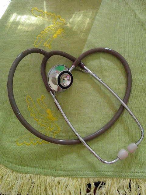
A stethoscope is a medical device for listening to sounds inside the body. The initial stethoscope was invented in the early 19th century by French physician Ren� Laennec, but was actually trying to achieve a rather different end: doctor-patient distance....
Showing posts with label procedures. Show all posts
Showing posts with label procedures. Show all posts
Sunday, June 22, 2014
Selection of Phlebotomy Venipuncture Site
Antecubital vein location varies slightly from person to person; however, two basic vein distribution arrangements referred to as the “H-shaped” and the “M-shaped” patterns are seen most often. The “H-shaped” pattern is so named because the most prominent veins in this pattern- the cephalic, cephalic median, median basilic, and basilic veins- are distributed on the arm in a way that resembles a slanted H. The most prominent veins of the M pattern- the cephalic, median cephalic, median basilic, and basilic veins- resemble the shape of an M. The H-shaped pattern is seen in approximately 70% of the population.
Factors in Vein Selection: Select the vein carefully. The brachial artery and several major nerves pass through the antecubital area. Accidental artery puncture and nerve injury are risks of venipuncture. Prioritizing veins can minimize the potential for accidental arterial puncture and nerve involvement. Typically, a tourniquet is used to aid in the selection of a vein unless specific tests require that a tourniquet not be used. A tourniquet is not necessary if veins are large and easily palpated. However, if only the basilic vein is visible without a tourniquet, one must be applied so the availability of safer veins (e.g. median and/or cephalic) can be assessed. Palpation is usually performed using the index finger. The collector’s thumb should not be used to palpate because it has a pulse beat. In addition to locating veins, the palpation pressure helps to differentiate veins from arteries, which pulsate, are more elastic, and have a thick wall.
Accidental Arterial Puncture: If during the procedure accidental arterial puncture is suspected (e.g. rapidly forming hematoma, rapid filling tube, and bright red blood), discontinue the venipuncture immediately. Remove the needle and apply direct forceful pressure to the puncture site for a minimum of 5 minutes until active bleeding has ceased. The nursing staff and physician must be notified and the incident documented according to institutional policy.
Consult with supervisory personnel to determine the suitability of the suspected arterial specimen for testing. If the specimen is acceptable it must be annotated that the specimen was an arterial specimen. In some cases different normal reference intervals are assigned to arterial blood. This information must be conveyed to the caregiver through Meditech Specimen Collection comment and Test Result comments.
Nerve Injury: If the patient feels a shooting, electric-like pain, or tingling or numbness proximal or distal to the venipuncture site, terminate the venipuncture and remove the needle immediately. Repeat the venipuncture in another site with a new sterile needle if needed. Document the incident and direct the patient to medical evaluation if indicated.
Monday, June 9, 2014
Supplies and Use of Supplies for Phlebotomy Venipuncture Procedure
Posted by
Channel Maymoon
Labels:
equipment,
nursing procedures,
nursing skills,
Phlebotomy,
procedures,
techniques
at
2:42 PM
By establishing a procedure for the correct collection of blood by venipuncture many pre-
analytical errors and patient management complications can be avoided. Patient safety is the ultimate goal above all other considerations. Cost, efficiency, etc are secondary to ensuring that in no way will the patient be harmed by the phlebotomy procedure. This includes all aspects of the procedure including ordering, drawing, labeling, handling and transporting the specimen. The quality of the patient results is directly dependent upon the quality of the specimen. By providing the highest standard of safety and quality of care customer service satisfaction can be achieved.
Supplies and use of supplies –(Refer to Standard Phlebotomy Tray policy)
1. Blood Collecting Trays
- Blood collecting trays should be lightweight and easy to handle with enough space and compartments for the various supplies.
2. Gloves
- Disposable latex, vinyl, polyethylene, or nitrile gloves provide barrier protection and must be worn for all venipuncture procedures to comply with OSHA regulations.
Latex free gloves must be worn for all patients with a hypersensitivity to latex proteins.
3. Hubs
–
a. All Vacutainer holders are to be SINGLE USE.
OSHA states “Blood tube holders, with needle attached, must be immediately discarded into an accessible sharps container after safety feature has been activated”. The re-use of vacutainer blood tube holders is strictly prohibited by OSHA and BVHS. (According to OSHA, “removing contaminated needles and re-using blood tube holders can expose workers to multiple hazards.”
b. Specimen transfer hubs are also available and, for our safety, are to be used before attempting to use a transfer needle. To use this device, simply attach it to the syringe and place the/each necessary vacutainer tube in the in the vacutainer holder until the appropriate amount of specimen is transferred.
4. Needles
- A large gauge (G) number indicates a small needle, while a small gauge number indicates alarge needle.
Needles must always be sterile and should never be recapped.
In order to prevent potential worker exposure, the needle safety feature should be activated immediately after specimen collection and discarded without disassembly into a sharps container. Needles are single use only.
a. BD Hypodermic needles
b. Butterfly “Push Button”
5. Sterile Syringes
-Sterile syringes must remain sterile. If removed from their container and not used immediately they are no longer considered sterile and are not to be used.
6. Blood Collection Tubes
- Venous blood collection tubes are manufactured to withdraw a predetermined volume of blood.
7. Tourniquets
-Tourniquets must be discarded immediately when contamination with blood or body fluids is obvious or suspected. Before drawing any in-patients, be sure to look around the room,
typically next to the sharps container, for a tourniquet that is specific for that patient. Out-patient draws and off-site tourniquets are replaced daily, or upon any sign of obvious or suspectedcontamination.
8. Antiseptics
- 70% isopropyl, PVP iodine prep pads, or 2% Iodine Tincture.
9. Gauze Pads
-Small, gauze pads should be available. Cotton balls may also be used.
10. Puncture-Resistant Disposable Container
-An approved puncture-resistant disposable container that is compliant with national or local regulations must be available for the disposal of the contaminated needle assembly. Such containers typically have a color regulated by each country, and a biohazard symbol.
11. Ice
-Ice or refrigerant should be available for specimens that require immediate chilling.
12. Bandages/Tape
- Adhesive bandages, preferably hypoallergenic, should be available, as well as gauze wraps for sensitive or fragile skin.
13. Warming Devices
- Warming devices may be used to dilate blood vessels and increase flow. When using commercial warmers follow the manufacturer’s recommendation. Warming devices should not exceed 42º C.
14. Specimen Collection Manual/Reference Lab Manual
- A test manual listing the tube(s) and volume requirements for various tests, specimen handling instructions, and precautions is available on all computers.
Wednesday, August 28, 2013
Electrocardiography: Equipment Preparation
Posted by
Channel Maymoon
Labels:
Cardiovascular Care,
ECGelectrocardiography,
electrocardiography,
equipment,
nursing,
preparation,
procedures
at
1:57 PM
One of the most valuable and frequently used diagnostic tools, electrocardiography (ECG) measures the heart's electrical activity as waveforms. Impulses moving through the heart's conduction system create electric currents that can be monitored on the body's surface. Electrodes attached to the skin can detect these electric currents and transmit them to an instrument that produces a record (the electrocardiogram) of cardiac activity.
ECG can be used to identify myocardial ischemia and infarction, rhythm and conduction disturbances, chamber enlargement, electrolyte imbalances, and drug toxicity.
The standard 12-lead ECG uses a series of electrodes placed on the extremities and the chest wall to assess the heart from 12 different views (leads). The 12 leads consist of three standard bipolar limb leads (designated I, II, III), three unipolar augmented leads (aVR, aVL, aVF), and six unipolar precordial leads (V1 to V6). The limb leads and augmented leads show the heart from the frontal plane. The precordial leads show the heart from the horizontal plane.
The ECG device measures and averages the differences between the electrical potential of the electrode sites for each lead and graphs them over time. This creates the standard ECG complex, called PQRST. The P wave represents atrial depolarization; the QRS complex, ventricular depolarization; and the T wave, ventricular repolarization. (See Reviewing ECG waveforms and components.)
Variations of standard ECG include exercise ECG (stress ECG) and ambulatory ECG (Holter monitoring). Exercise ECG monitors heart rate, blood pressure, and ECG waveforms as the patient walks on a treadmill or pedals a stationary bicycle. For ambulatory ECG, the patient wears a portable Holter monitor to record heart activity continually over 24 hours.
Today, ECG is typically accomplished using a multichannel method. All electrodes are attached to the patient at once, and the machine prints a simultaneous view of all leads.
Today, ECG is typically accomplished using a multichannel method. All electrodes are attached to the patient at once, and the machine prints a simultaneous view of all leads.
Equipment
ECG machine ; recording paper ; disposable pregelled electrodes ; 4″ × 4″ gauze pads ; optional: clippers, marking pen.
ECG machine ; recording paper ; disposable pregelled electrodes ; 4″ × 4″ gauze pads ; optional: clippers, marking pen.
Preparation of equipment
Place the ECG machine close to the patient's bed, and plug the power cord into the wall outlet. If the patient is already connected to a cardiac monitor, remove the electrodes to accommodate the precordial leads and minimize electrical interference on the ECG tracing. Keep the patient away from objects that might cause electrical interference, such as equipment, fixtures, and power cords.
Saturday, August 24, 2013
Basic Procedures That Must Be Understood By Every Nurse
Patients come to the hospital and other health facilities because they require skilled clinical observation and treatment. Millions of people hospitalized each year, and for the most part, it was a trying experience. Inpatient care dealing with patients' needs for privacy and control of his life. He should release at least part of the normal routine. He had to rely on you and your co-workers to meet basic needs. Depending on the complexity of health problems, he and his family may also require teaching, counseling, coordination of care, development of community support systems, and help in coping with changes related to health in his life.
Some broader aims of your care are helping the patient cope with restricted mobility; giving him a comfortable, stimulating environment; making sure his stay is free from hazards; promoting an uneventful recovery; and helping him return to his normal life.
Each time the patient's condition deter or prevent mobility, then your nursing goals include promoting independence by motivating him, helped him set goals, to prevent injury and complications of immobility, he teaches the skills needed, and encourage a positive body image, especially if he faces a long term or permanent immobility.
Besides weakening the patient, illness and any accompanying treatment may impair his judgment and contribute to accidents. Be alert to hazards in the patient's environment, and teach him and his family to recognize and correct them. When caring for a patient with restricted mobility, you must help him as he's moved, lifted, and transported. By using proper body mechanics and appropriate assistive devices, you can prevent injury, fatigue, and discomfort for the patient and yourself. To prevent complications, be sure to use correct positioning, meticulous skin care, assistive devices, and regular turning and range-of-motion exercises.
The first step toward rehabilitation typically is progressive ambulation, which should begin as soon as possible if necessary, using such assistive devices as a cane, crutches, or a walker. Demonstrating a technique such as transferring from a bed to a wheelchair during hospitalization helps the patient and his family to understand it. Allowing them to practice it under your supervision gives them the confidence to perform it at home. Encourage them to provide positive reinforcement to motivate the patient to work toward his goals.
Subscribe to:
Comments (Atom)
Powered by Blogger.


