• How Stethoscope Works....

    A stethoscope is a medical device for listening to sounds inside the body. The initial stethoscope was invented in the early 19th century by French physician Ren� Laennec, but was actually trying to achieve a rather different end: doctor-patient distance....

  • Care Plan For Decreased Cardiac Output...

    Nursing diagnosis for decreased cardiac output may be related to altered myocardial contractility, inotropic changes; alterations in rate, rhythm, electrical conduction; or structural changes, such as valvular defects and ventricular aneurysm. ...

  • ECG Waveforms And Components

    The electrocardiogram (ECG) is a graphic recording ofelectric potentials generated by the heart.The signals are detected by means of metal electrodes attached to the extremities and chest wall and are then amplified and recorded by the electrocardiograph. ECG leads actually display the instantaneous differences in potential between these electrodes. ...

Saturday, November 30, 2013

Nurse’s Ethical Duty in Wound Care

Performing an evaluation, assessment, or management of any type of wound is an ethical endeavor and may present ethical challenges at times. The specific concepts of paternalism, autonomy, beneficence, nonmaleficence, fidelity, role fidelity, veracity, therapeutic privilege, conflict of interest, confidentiality, and justice will be addressed.
The nurse might wonder why consideration of morals is of any importance when what he or she is doing is providing clinical services for some type of wound. The practice of wound care is fraught with areas in which the morals or society’s determination of right and good conduct of the health-care professional may be seriously tested. Understanding the concepts of morals, moral duty, and moral obligation are critical in providing wound care.
Specific obligations and duties for the privilege of professional access to patients, including the following:
  • First, the patient’s interests are placed above the personal interest of the nurse. If this duty is overlooked or forgotten, the contract (standard of practice) among the health-care provider, the health-care organization, and the patient is broken. — Example: The health-care provider conducts a seminar and needs wound photographs to supplement the written and verbal components of the presentation. The provider takes photographs of the patient’s wounds solely for the purpose of using them in the seminar. The only reason for taking these photographs is for the convenience of the health-care provider, and therefore the activity is actually for the nurse’s personal interest and not for the patient’s best interest. The patient would need to grant the nurse informed consent to use the photographs to avoid any consideration that the photographs are for personal interest. The nurse would need to assure the patient that any refusals on the patient’s part would have no effect on the nurse-patient relationship or the patient’s treatment.
  • The patient’s privacy is protected from another individual’s or society’s desire to know details of the patient’s treatment. It is the health-care provider’s responsibility to have a complete understanding of the legal rights of all involved. It is ultimately the responsibility of the nurse to know the legal rights of the patient, family, and health-care provider. However, in many areas of the world, the general public does not have any legal right to knowledge concerning the patient’s care, progress, or prognosis. The health-care provider must identify if the health-care organization has a policy or procedure concerning this challenge.
  • Does the health-care provider have a duty to treat the patient who has a wound(s)?
It is important to remember that this information concerns the ethical decision making only and is not to be construed as presenting a legal argument for or against treatment. Failure to treat may have potential legal consequences.
Read More

Thursday, November 28, 2013

Overview of Respiratory Function

The major function of lungs and pulmonary circulation as the pulmonary system is to deliver oxygen (O2) to cells and remove carbon dioxide (CO2) from the cells (gas exchange). The adequacy of oxygenation and ventilation is measured by partial pressure of arterial oxygen (PaO2) and partial pressure of arterial carbon dioxide (PaCO2). The pulmonary system also functions as a blood reservoir for the left ventricle when it is needed to boost cardiac output; as a protector for the systemic circulation by filtering debris/particles; as a fluid regulator so water can be kept away from alveoli; and as a provider of metabolic functions such as surfactant production and endocrine functions.
Terminology in Respiratory
  • Alveolus—air sac where gas exchange takes place
  • Apex—top portion of the upper lobes of lungs
  • Base—bottom portion of lower lobes of lungs, located just above the diaphragm
  • Bronchoconstriction—constriction of smooth muscle surrounding bronchioles
  • Bronchus—large airways; lung divides into right and left bronchi
  • Carina—location of division of the right and left main stem bronchi
  • Cilia—hairlike projections on the tracheobronchial epithelium, which aid in the movement of secretions and removal of debris
  • Compliance—ability of the lungs to distend and change in volume relative to an applied change in pressure (eg, emphysema—lungs very compliant; fibrosis—lungs noncompliant or stiff)
  • Dead space—ventilation that does not participate in gas exchange; also known as wasted ventilation when there is adequate ventilation but no perfusion, as in pulmonary embolus or pulmonary vascular bed occlusion. Normal dead space is 150 mL.
  • Diaphragm—primary muscle used for respiration; located just below the lung bases, it separates the chest and abdominal cavities
  • Diffusion (of gas)—movement of gas from area of higher to lower concentration
  • Dyspnea—subjective sensation of breathlessness associated with discomfort, often caused by a dissociation between motor command and mechanical response of the respiratory system as in:
    • Respiratory muscle abnormalities (hyperinflation and airflow limitation from chronic obstructive pulmonary disease [COPD]).
    • Abnormal ventilatory impedance (narrowing airways and respiratory impedance from COPD or asthma).
    • Abnormal breathing patterns (severe exercise, pulmonary congestion or edema, recurrent pulmonary emboli).
    • Arterial blood gas (ABG) abnormalities (hypoxemia, hypercarbia).
  • Hemoptysis—coughing up of blood
  • Hypoxemia—PaO2 less than normal, which may or may not cause symptoms (Normal PaO2 is 80 to 100 mm Hg on room air.)
  • Hypoxia—insufficient oxygenation at the cellular level due to an imbalance in oxygen delivery and oxygen consumption (Usually causes symptoms reflecting decreased oxygen reaching the brain and heart.)
  • Mediastinum—compartment between lungs containing lymph and vascular tissue that separates left from right lung
  • Orthopnea—shortness of breath when in reclining position
  • Paroxysmal nocturnal dyspnea—sudden shortness of breath associated with sleeping in recumbent position
  • Perfusion—blood flow, carrying oxygen and CO2 that passes by alveoli
  • Pleura—serous membrane enclosing the lung; comprised of visceral pleura, covering all lung surfaces, and parietal pleura, covering chest wall and mediastinal structures, between which exists a potential space
  • Pulmonary circulation—network of vessels that supply oxygenated blood to and remove CO2-laden blood from the lungs
  • Respiration—inhalation and exhalation; at the cellular level, a process involving uptake of oxygen and removal of CO2 and other products of oxidation
  • Shunt—adequate perfusion without ventilation, with deoxygenated blood conducted into the systemic circulation, as in pulmonary edema, atelectasis, pneumonia, COPD
  • Surfactant—fluid secreted by alveolar cells that reduces surface tension of pulmonary fluids and aids in elasticity of pulmonary tissue
  • Ventilation—movement of air (gases) in and out of the lungs
  • Ventilation-perfusion (V/Q) imbalance or mismatch—imbalance of ventilation and perfusion; a cause for hypoxemia. V/Q mismatch can be due to:
    • Blood perfusing an area of the lung where ventilation is reduced or absent.
    • Ventilation of parts of lung that are not perfused.
Read More

Wednesday, November 27, 2013

Biologic and Genetic Principles on Nursing

The impact of genetics on nursing is significant. The American Nurses Association (ANA) officially recognized genetics as a nursing specialty. This effort was spearheaded by the International Society of Nurses in Genetics (ISONG), which also initiated credentialing for the Advanced Practice Nurse in Genetics and the Genetics Clinical Nurse. ANA and ISONG have collaborated in the establishment of a scope and standards of practice for nurses in genetics practice. Essential Nursing Competencies and Curricula Guidelines for Genetics and Genomics were finalized in 2006. They reflect the minimal genetic and genomic competencies for every nurse regardless of academic preparation, practice setting, role, or specialty.

Cell as The Basic Unit of Biology
  • Cytoplasm—contains functional structures important to cellular functioning, including mitochondria, which contain extranuclear deoxyribonucleic acid (DNA) important to mitochondrial functioning.
  • Nucleus—contains 46 chromosomes in each somatic (body) cell, or 23 chromosomes in each germ cell (egg or sperm).
Chromosomes
Each somatic cell with a nucleus has 22 pairs of autosomes (the same in both sexes) and 1 pair of sex chromosomes. Females have two X sex chromosomes; males have one Y sex chromosome and one X sex chromosome. Normally, at conception, each individual receives one copy of each chromosome from the maternal egg cell (1 genome) and one copy of each chromosome from the paternal sperm cell (1 genome), for a total of 46 chromosomes (2 genomes). Karyotype is the term used to define the chromosomal complement of an individual, for example, 46, XY, as is determined by laboratory chromosome analysis. Each chromosome contains 800 to 3,000 genes.
 
Genes
Gene is the basic unit of inherited information. Each copy of the human genome in the nucleus has about 30,000 genes. Cells also have some nonnuclear genes located within the mitochondria within the cytoplasm. Alternate forms of a gene are termed alleles. For each gene, an individual receives one allele from each parent, and thus has two alleles for each gene on the autosomes and also on the X chromosomes in females. Males have only one X chromosome and, therefore, have only one allele for all genes on the X chromosome; they are hemizygous for all X-linked genes. At any autosomal locus, or gene site, an individual can have two identical alleles (homozygous) for that locus or can have two different alleles (heterozygous) at a particular locus. Genotype refers to the constitution of the genetic material of an individual; for practical purposes it is commonly used to address a specific gene pair. For example, the gene for sickle cell disease, the gene for cystic fibrosis, or the gene for familial polyposis. Phenotype refers to the physical or biochemical characteristics an individual manifests regarding expression of the presence of a particular feature, or set of features, associated with a particular gene. Each gene is composed of a unique sequence of DNA bases.
 
DNA: Nuclear and Mitochondrial
  • Human DNA is a double-stranded helical structure comprised of four different bases, the sequence of which codes for the assembly of amino acids to make a protein—for example, an enzyme. These proteins are important for the following reasons:
    • For body characteristics such as eye color.
    • For biochemical processes such as the gene for the enzyme that digests phenylalanine.
    • For body structure such as a collagen gene important to bone formation.
    • For cellular functioning such as genes associated with the cell cycle.
  • The four DNA bases are adenine, guanine, cytosine, and thymine-A, G, C, and T.
  • A change, or mutation, in the coding sequence, such as a duplicated or deleted region, or even a change in only one base, can alter the production or functioning of the gene or gene product, thus affecting cellular processes, growth, and development.
  • DNA analysis can be done on almost any body tissue (blood, muscle, skin) using molecular techniques (not visible under a microscope) for mutation analysis of a specific gene with a known sequence or for DNA linkage of genetic markers associated with a particular gene.
Normal Cell Division
Mitosis occurs in all somatic cells, which, under normal circumstances, results in the formation of cells identical to the original cell with the same 46 chromosomes.
Meiosis, or reduction division, occurs in the germ cell line, resulting in gametes (egg and sperm cells) with only 23 chromosomes, one representative of each chromosome pair.
During the process of meiosis, parental homologous chromosomes (from the same pair) pair and undergo exchanges of genetic material, resulting in recombinations of alleles on a chromosome and thus variation in individuals from generation to generation.
Read More

Monday, November 25, 2013

Using Electrocardiography (ECG) to Measures the Heart's Electrical Activity

Prepare the machine by placing the ECG machine close to the patient's bed, and plug the power cord into the wall outlet. To accommodate the precordial leads and minimize electrical interference on the ECG tracing, remove the electrodes if the patient is already connected to a cardiac monitor. Keep the patient away from objects that might cause electrical interference, such as equipment, fixtures, and power cords.

Explain the procedure to the patient as you set up the machine to record a 12-lead ECG. Tell him that the test records the heart's electrical activity and it may be repeated at certain intervals. Also, tell him that the test typically takes about 5 minutes. Emphasize that no electrical current will enter his body.

Have the patient lie in a supine position in the center of the bed with his arms at his sides. You may raise the head of the bed to promote his comfort. Expose his arms and legs, and drape him appropriately. His arms and legs should be relaxed to minimize muscle trembling, which can cause electrical interference.
Place the patient's hands under his buttocks to prevent muscle tension if the bed is too narrow. Also use this technique if the patient is shivering or trembling. Make sure his feet aren't touching the bed board.
Select flat, fleshy areas to place the electrodes. Avoid muscular and bony areas. If the patient has an amputated limb, choose a site on the stump. If an area is excessively hairy, clip it. Clean excess oil or other substances from the skin to enhance electrode contact.
Apply the electrode paste or gel or the disposable electrodes to the patient's wrists and to the medial aspects of his ankles. If you're using paste or gel, rub it into the skin. If you're using disposable electrodes, peel off the contact paper and apply them directly to the prepared site, as recommended by the manufacturer's instructions. To guarantee the best connection to the leadwire, position disposable electrodes on the legs with the lead connection pointing superiorly.
If you're using paste or gel, secure electrodes promptly after you apply the conductive medium. This prevents drying of the medium, which could impair ECG quality. Never use alcohol or acetone pads in place of the electrode paste or gel because they impair electrode contact with the skin and diminish the transmission quality of electrical impulses.
Connect the limb leadwires to the electrodes. Make sure the metal parts of the electrodes are clean and bright. Dirty or corroded electrodes prevent a good electrical connection.
You'll see that the tip of each leadwire is lettered and color-coded for easy identification. The white or RA leadwire goes to the right arm; the green or RL leadwire, to the right leg; the red or LL leadwire, to the left leg; the black or LA leadwire, to the left arm; and the brown or V1 to V6 leadwires, to the chest.
Now, expose the patient's chest. Put a small amount of electrode gel or paste on a disposable electrode at each electrode position.
If your patient is a woman, be sure to place the chest electrodes below the breast tissue. In a large-breasted woman, you may need to displace the breast tissue laterally.
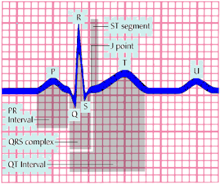
Check to see that the paper speed selector is set to the standard 25 mm/second and that the machine is set to full voltage. The machine will record a normal standardization mark—a square that's the height of two large squares or 10 small squares on the recording paper. Then, if necessary, enter the appropriate patient identification data.
If any part of the waveform extends beyond the paper when you record the ECG, adjust the normal standardization to half-standardization. Note this adjustment on the ECG strip because this will need to be considered in interpreting the results.
Now you're ready to begin the recording. Ask the patient to relax and breathe normally. Tell him to lie still and not to talk when you record his ECG. Then press the AUTO button. Observe the tracing quality. The machine will record all 12 leads automatically, recording three consecutive leads simultaneously. Some machines have a display screen so you can preview waveforms before the machine records them on paper.
When the machine finishes recording the 12-lead ECG, remove the electrodes and clean the patient's skin. After disconnecting the leadwires from the electrodes, dispose of or clean the electrodes, as indicated.
 
 
Read More
Powered by Blogger.

Search This Blog