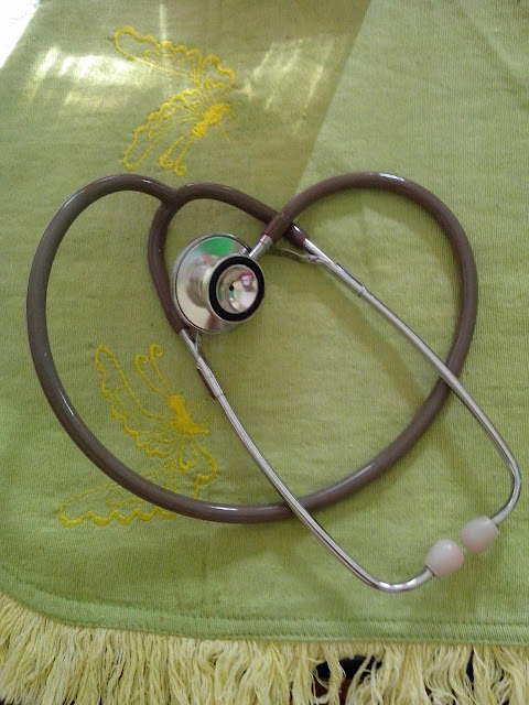
A stethoscope is a medical device for listening to sounds inside the body. The initial stethoscope was invented in the early 19th century by French physician Ren� Laennec, but was actually trying to achieve a rather different end: doctor-patient distance....
Showing posts with label nerve. Show all posts
Showing posts with label nerve. Show all posts
Friday, July 10, 2015
Assessment of Cranial Nerves in The Child
Assessment of cranial nerves in the child. Obtained from Hadley (1994). S Sensory, M motor, EOM extraocular movement.
Cranial
|
Test for function
|
|---|---|
I Olfactory (S)
|
|
Olfactory nerve, mucous membrane of nasal passages and turbinates |
With eyes closed child is asked to identify familiar odors such as peanut butter, orange, and peppermint. Test each nostril separately |
II Optic (S)
|
|
Optic nerve, retinal rods and cones |
Check visual acuity, peripheral vision, color vision, perception of light in infants, fundoscopic examination for normal optic disk |
III Oculomotor (M)
|
|
Muscles of the eyes (superior rectus, inferior rectus, medial rectus, inferior oblique) |
Have child follow an object or light with the eyes (EOM) while head remains stationary. Check symmetry of corneal light reflex. Check for nystagamus (direction elicited, vertical, horizontal, rotary). Check cover-uncover test. |
Muscles of iris and ciliary body |
Reaction of pupils so light, both direct and consensual, accommodation |
Levator palpebral muscle |
Check for symmetric movement of upper eyelids. Note ptosis |
IV Trochlear (M)
|
|
Muscles of eye (superior oblique) |
Check the range of motion of the eyes downward (EOM). Check for nystagmus |
V Trigeminal (M, S)
|
|
Muscles of mastication (M) |
Palpate the child’s jaws, jaw muscles, and temporal muscles for strength and symmetry. Ask child to move lower jaw from side to side against resistance of the examiner’s hand |
Sensory innervation of face (S) |
Test child for sensation using a wisp of cotton, warm and cold water in test tubes, and a sharp object on the forehead, cheeks, and jaw. Check corneal reflex by touching a wisp of cotton to each cornea. The normal response is blink |
VI Abducens (M)
|
|
Muscles of eye (lateral rectus) |
Have child look to each side (EOM) |
VII Facial (M, S)
|
|
Muscles for facial expression |
Have child make faces: look at the ceiling, frown, wrinkle forehead, blow out cheeks, smile. Check for strength, asymmetry, paralysis |
Sense of taste on anterior two-thirds of tongue. Sensation of external ear canal, lachrymal, submaxillary, and sublingual glands |
Have a child identify salt, sugar, bitter (flavoring extract), and sour substances by placing substance on anterior sides of tongue. Keep tongue out until substance is identified. Rinse mouth between substances |
VIII Acoustic (S)
|
|
Equilibrium (vestibular nerve) |
Note equilibrium or presence of vertigo (Romberg sign) |
Auditory acuity (cochlear nerve) |
Test hearing. Use a tuning fork for the Weber and Rinne tests. Test by whispering and use of a watch |
IX Glossopharyngeal (M, S)
|
|
Pharynx, tongue (M) |
Check elevation of palate with “ah” or crying. Check for movement and symmetry. Stimulate posterior pharynx for gag reflex |
Sense of taste posterior third of the tongue |
Test sense of taste on posterior portion of tongue |
X Vagus (M, S)
|
|
Mucous membrane of pharynx, larynx, bronchi, lungs, heart, esophagus, stomach, and kidneys Posterior surface of external ear and external auditory meatus |
Note same as for glossopharyngeal. Note any hoarseness or stridor. Check uvula for midline position and movement with phonation. Stimulate uvula on each side with tongue depressor – should rise and deviate to stimulated side. Check gag reflex. Observe ability to swallow |
XI Accessory (M)
|
|
Sternocleidomastoid and upper trapezius muscles |
Have child shrug shoulders against mild resistance. Have child turn head to one side against resistance of examiner’s hand. Repeat on the other side. Inspect and palpate muscle strength, symmetry for both maneuvers |
XII Hypoglossal (M)
|
|
Muscle of tongue |
Have child move the tongue in all directions, then stick out tongue as far as possible: check for tremors or deviations. Test strength by having child push tongue against inside cheek against resistance on outer cheek. Note strength, movement, symmetry |
Wednesday, July 8, 2015
Cranial Nerve - Brainstem Function
Cranial nerve assessment is basically an assessment of brainstem function because nuclei of 10 of the 12 cranial nerves are located in the brainstem. The proximity of these nuclei to the reticular activating system (arousal center) located in the midbrain is the anatomic rationale for assessing cranial nerves in conjunction with LOC. Important neurological functions and protective reflexes are mediated by the cranial nerves and many functions are dependent on more than one nerve. Some of the cranial nerves have both motor and sensory functions.
The two cranial nerves that do not arise in the brainstem are the olfactory nerve (CN I) and the optic nerve (CN II). CN I is located in the medial frontal lobe and is responsible for the sense of smell. This can be difficult to assess in the younger child, so is often omitted unless there is specific concern that there has been damage in that area. Taste may also be affected with injuries to CN I. CN II is assessed by determining a child’s visual acuity. This may be done more formally with visual screening or more generally by noting if the child’s vision appears normal in routine activities.
Pupil size and response to direct light are mediated by CN II and the oculomotor nerve (CN III) as well as the sympathetic nervous system. Many things can affect the pupillary response in a child, including damage to the eye or the cranial nerves, pressure on the upper brainstem, local and systemic effects of certain drugs, anoxia, and seizures. Pupillary size varies with age and is determined by the amount of sympathetic input, which dilates the pupil and is balanced by the parasympathetic input on CN III, which constricts the pupil. Pupillary response in the eye that is being checked with direct light as well as the other pupil (consensual response) are significant in that they can point to where damage to nerves exists and are an objective clinical sign that can be followed over time .
 |
| Diagram of the base of the brain showing entrance and exits of the cranial nerves |
The two cranial nerves that do not arise in the brainstem are the olfactory nerve (CN I) and the optic nerve (CN II). CN I is located in the medial frontal lobe and is responsible for the sense of smell. This can be difficult to assess in the younger child, so is often omitted unless there is specific concern that there has been damage in that area. Taste may also be affected with injuries to CN I. CN II is assessed by determining a child’s visual acuity. This may be done more formally with visual screening or more generally by noting if the child’s vision appears normal in routine activities.
Pupil size and response to direct light are mediated by CN II and the oculomotor nerve (CN III) as well as the sympathetic nervous system. Many things can affect the pupillary response in a child, including damage to the eye or the cranial nerves, pressure on the upper brainstem, local and systemic effects of certain drugs, anoxia, and seizures. Pupillary size varies with age and is determined by the amount of sympathetic input, which dilates the pupil and is balanced by the parasympathetic input on CN III, which constricts the pupil. Pupillary response in the eye that is being checked with direct light as well as the other pupil (consensual response) are significant in that they can point to where damage to nerves exists and are an objective clinical sign that can be followed over time .
Subscribe to:
Comments (Atom)
Powered by Blogger.


