• How Stethoscope Works....

    A stethoscope is a medical device for listening to sounds inside the body. The initial stethoscope was invented in the early 19th century by French physician Ren� Laennec, but was actually trying to achieve a rather different end: doctor-patient distance....

  • Care Plan For Decreased Cardiac Output...

    Nursing diagnosis for decreased cardiac output may be related to altered myocardial contractility, inotropic changes; alterations in rate, rhythm, electrical conduction; or structural changes, such as valvular defects and ventricular aneurysm. ...

  • ECG Waveforms And Components

    The electrocardiogram (ECG) is a graphic recording ofelectric potentials generated by the heart.The signals are detected by means of metal electrodes attached to the extremities and chest wall and are then amplified and recorded by the electrocardiograph. ECG leads actually display the instantaneous differences in potential between these electrodes. ...

Friday, September 13, 2013

Buying The Right Parts For Your Vehicle

Before you go shopping for some parts to replace those on your vehicle, read the tips in this section carefully. They can help you avoid what’s probably the most annoying part of any automotive job: disabling your vehicle to work on it only to find that you need it to drive back to the store to exchange the stuff they sold you in error!

To buy the proper parts for your vehicle, you must know its specifications (or “specs,” as they’re often called). Most of this information should be in your owner’s manual, and a lot of it is also printed on metal tags or decals located inside your hood. You can usually find these in front of the radiator, inside the fenders, on the inside of the hood — anywhere the auto manufacturer thinks you’ll find them. I know of one car that has its decal inside the lid of the glove compartment. These ID tags also provide a lot of other information about where the vehicle was made, what kind of paint it has, and so on.

The service manual for your vehicle should have the specs for the parts you need, and the parts department at your dealership or a reputable auto supply store can also look them up for you.

It’s a good idea to stick with parts from the same manufacturer as those that your vehicle originally came with. That brand may be listed in a service manual for your vehicle. If you don’t have a service manual, tell the sales clerk at the auto parts store that you want OEM (original equipment manufacturer) parts. Quality aftermarket parts are available as well, but unless you trust your parts seller’s recommendations, or you’ve already used a particular aftermarket brand and had good luck with it, stick with OEM parts.

If you can’t find specs for buying and gapping spark plugs in your owner’s or service manual or on your vehicle, just ask to the expert. 

When you go to buy parts, keep in mind that most professional mechanics get discounts at auto parts stores. Ask if you can get a discount given that you’re installing the parts yourself. It can’t hurt to try. Even if you don’t get a price break on parts, you’ll still be ahead of the game because you won’t have to pay labor charges.

 
Read More

Saturday, September 7, 2013

How a Stethoscope Works

A stethoscope is a medical device for listening to sounds inside the body. The initial stethoscope was invented in the early 19th century by French physician René Laennec, but was actually trying to achieve a rather different end: doctor-patient distance. The stethoscope can be placed against the patient's chest to listen to her breath or heartbeat, or against the lower abdomen to listen to the intestines. On one end of the stethoscope is a diaphragm, a vibrating membrane designed to pick up sound. The diaphragm is connected to a hollow, air-filled tube. That tube splits in two and leads to earpieces, which the doctor wears.

When the heart beats or the lung fills with air, it produces small sound vibrations through the body. These vibrations are picked up and amplified by the diaphragm. The sound passes into the tube, which transfers it into the doctor's earpieces. There are also electrical stethoscopes, which use a kind of microphone to pick up and amplify the sound.

To understand how a stethoscope conveys, say, the "lub-lub" of a heart beat from a heart to a doctor's ears, we'll start with the core components of the tool.
 
Chestpiece: This is the part that contacts the patient, capturing sound. The chestpiece is constructed of metal and is typically double-sided to work for differently sized patients and different areas of the body. The chestpiece has deep cups that capture sounds from the target area and may be ringed with a "chill ring" to keep the patient from being uncomfortable when touched by the cold metal chestpiece. There are two sides of the chestpiece. On one side is the diaphragm, a flat, metal disc that in turn contains a flat, plastic disc. The diaphragm is the larger component of the chestpiece. On the other side is the bell, a hollow, bell-shaped piece of metal with a tiny hole on top. The bell is better at picking up low-pitch sounds, such as heart murmurs (the aforementioned "whoosh"); the diaphragm excels in the higher-pitch range, which includes normal breath sounds and heartbeats ("lub-lub").
 
Acoustic Tubes and Ear Tubes: A Y-shaped configuration of rubber tubesruns from the chestpiece to the headset. The sounds picked up by the chestpiece initially travel through a single tube, eventually splitting into two channels as they near the headset so the listener can hear it in both ears. Stethoscope tubing typically ranges from about 18 to 27 inches (45 to 68 centimeters) long. The acoustic tube connects the ear tubes to the chestpiece and is a flexible tube made of a rubberized material. The acoustic tubing can be filled with different materials to best transmit sound from the patient to the health-care provider. The ear tubes attach the earpieces to the main body of the stethoscope. They are typically constructed of metal and consist of hollow tubes that are ideally suited to transmitting low-frequency sound.
 
Earpieces/ Headset: The earpieces are fitted into the ears and are typically rubberized for comfort and to isolate from external sounds. They are designed to transmit sounds directly into the ears. The rubber tubing ends at a set of metal tubes that carry the sound to the eartips in the listener's ears. The eartips are made of soft rubber, not only for comfort but also to create a seal that helps block out environmental noise.
 
 
Read More

Friday, September 6, 2013

Essential Skills For Assessment In Nursing Process Steps

Assessment requires the use of the skills needed for interviewing, conducting a physical examination, and observing patients. As with the nursing process itself, these skills are not used one at a time. While you are interviewing the patient, you are also observing and determining physical areas that require a detailed physical assessment. While completing a physical assessment, you are asking questions (interviewing) and observing the patient’s physical appearance as well as the patient’s response to the physical examination.

Interviewing generally starts with gathering data for the nursing history. In this interview, you ask for general demographic information such as name, address, date of last hospitalization, age, allergies, current medications, and the reason the patient was admitted. Depending on the agency’s admission form, you may then progress to other specific questions or a physical assessment.

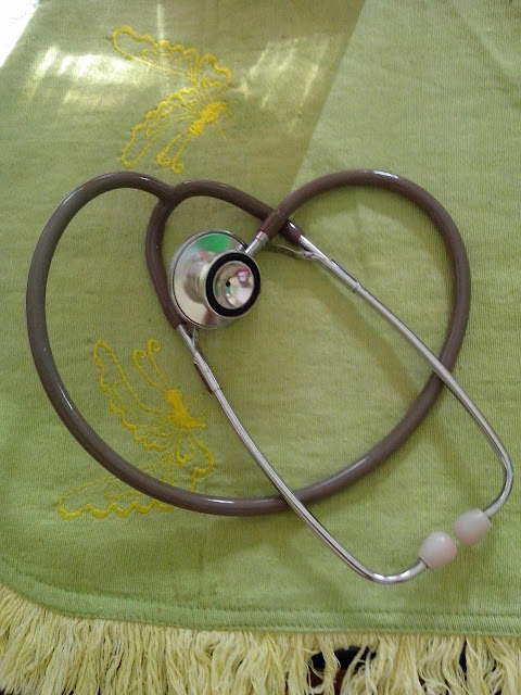
The physical assessment calls for four skills: inspection, palpation, percussion, and auscultation. Inspection means careful and systematic observation throughout the physical examination, such as observation for and recording of any skin lesions. Palpation is assessment by feeling and touching the patient. Assessing the differences in temperature between a patient’s upper and lower arm would be an example of palpation. Another common example of palpation is breast self-examination. Percussion involves touching, tapping, and listening. Percussion allows determination of the size, density, locations, and boundaries of the organs. Percussion is usually performed by placing the index or middle finger of one hand firmly on the skin and striking with the middle finger of the other hand. The resultant sound is dull if the body is solid under the fingers (such as at the location of the liver) and hollow if there is a body cavity under the finger (such as at the location of the abdominal cavity). Auscultation involves listening with a stethoscope and is used to help assess respiratory, circulatory, and gastrointestinal status.

The physical assessment may be performed using a head-to-toe approach, a body system approach, or a functional health pattern approach. In the head-to-toe approach, you begin with the patient’s general appearance and vital signs. You then progress, as the name indicates, from the head to the extremities.

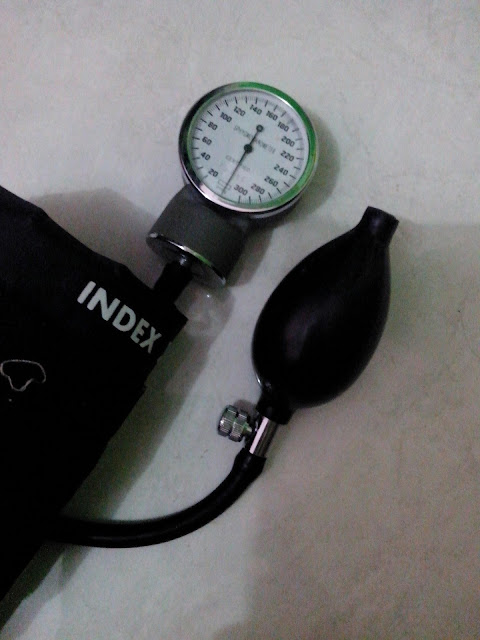
The body system approach to physical assessment focuses on the major body systems. As the nurse is conducting the nursing history interview, she or he will get a firm idea of which body systems need detailed examination. An example is a cardiovascular examination, where the apical and radial pulses, blood pressure (BP), point of maximum intensity (PMI), heart sounds, and peripheral pulses are examined.

The functional health pattern approach is based on Gordon’s Functional Health Patterns typology and allows the collection of all types of data according to each pattern. This is the approach used by this book and leads to three levels of assessment. First is the overall admission assessment, where each pattern is assessed through the collection of objective and subjective data. This assessment indicates patterns that need further attention, which requires implementation of the second level of pattern assessment. The second level of pattern assessment indicates which nursing diagnoses within the pattern might be pertinent to this patient, which leads to the third level of assessment, the defining characteristics for each individual nursing diagnosis. Having a three-tiered assessment might seem complicated, but each assessment is so closely related that completion of the assessment is easy. A primary advantage in using this type of assessment is the validation it gives to the nurse that the resulting nursing diagnosis is the most correct diagnosis. Another benefit to using this type of assessment is that grouping of data is already accomplished and does not have to be a separate step.

 
Read More

Care Plan Or Planning Of Care?

Revisions of nursing standards created questions regarding the necessity of nursing care plans. Some have predicted the rapid demise of the care plan, according to Brider, but review of the revised nursing standards shows that the standards require not less but more detailed care planning documentation in the patient’s medical record.

Review of the new criteria indicates that the standards require documentation related to the nursing process. For example, the plan of care statement reads:

A plan, based on data gathering during patient assessment, that identifies the patient’s care needs, tests the strategy for providing services to meet those needs, documents treatment goals or objectives, outlines the criteria for terminating specified interventions, and documents the individual’s progress in meeting specified goals and objectives. The format of the “plan” in some organizations may be guided by patient-specific policies and procedures, protocols, practice guidelines, clinical paths, care maps, or a combination of these. The plan of care may include care, treatment, habilitation and rehabilitation.

Rather than eliminating care plans, the requirements call for a more specific as well as a more permanent documentation of the plan of care. This documentation must be in the medical record. The standard indicates that a separate care plan form is no longer necessary; however, the standard also still allows a separate care plan form. Various institutions are now testing flexible ways of documenting care planning. The care plan is not dead; rather, it is revised to more clearly reflect the important role of nursing in the patient’s care. No longer a separate, often discarded, and irrelevant page, the plan of care must be part of the permanent record. The flow sheets developed for this book offer guidelines for computerizing information regarding nursing care.

Faculty can use the revised standards to assist students in developing expertise beyond writing extensive nursing care plans. This additional expertise requires the new graduate to integrate all phases of the nursing process into the permanent record. Rather than eliminating the need for care planning and nursing diagnosis, the standards have reinforced the importance of nursing care and nursing diagnosis.

 
Read More

Care Plan For Decreased Cardiac Output

Nursing diagnosis for decreased cardiac output may be related to altered myocardial contractility, inotropic changes; alterations in rate, rhythm, electrical conduction; or structural changes, such as valvular defects and ventricular aneurysm.

It is possibly evidenced by increased heart rate (tachycardia), dysrhythmias, ECG changes; changes in BP (hypotension, hypertension); extra heart sounds (S3, S4); decreased urine output; diminished peripheral pulses; cool, ashen skin and diaphoresis; orthopnea, crackles, JVD, liver engorgement, edema; or chest pain

Desired outcomes for this nursing diagnosis are, client will have Cardiac Pump Effectiveness-NOC by evaluation criteria

  • Display vital signs within acceptable limits, dysrhythmias absent or controlled, and no symptoms of failure, for example, hemodynamic parameters within acceptable limits and urinary output adequate.
  • Report decreased episodes of dyspnea and angina.

Client also will have Cardiac Disease Self-Management-NOC by evaluation criteria Participate in activities that reduce cardiac workload.

Possible intervention : Hemodynamic Regulation-NIC by action such as

  • Auscultate apical pulse; assess heart rate, rhythm, and document dysrhythmia if telemetry available. Tachycardia is usually present, even at rest, to compensate for decreased ventricular contractility. Premature atrial contractions (PACs), paroxysmal atrial tachycardia (PAT), PVCs, multifocal atrial tachycardia (MAT), and AF are common dysrhythmias associated with HF, although others may also occur. Note: Intractable ventricular dysrhythmias unresponsive to medication suggest ventricular aneurysm.
  • Note the heart sounds. S1 and S2 may be weak because of diminished pumping action. Gallop rhythms are common (S3 and S4), produced as blood flows into noncompliant, distended chambers.
  • Palpate peripheral pulses. Decreased cardiac output may be reflected in diminished radial, popliteal, dorsalis pedis, and post-tibial pulses. Pulses may be fleeting or irregular to palpation, and pulsus alternans may be present.
  • Inspect skin for pallor and cyanosis. Pallor is indicative of diminished peripheral perfusion secondary to inadequate cardiac output, vasoconstriction, and anemia. Cyanosis may develop in refractory HF. Dependent
    areas are often blue or mottled as venous congestion increases.
  • Monitor urine output, noting decreasing output and dark or concentrated urine. Kidneys respond to reduced cardiac output by retaining water and sodium. Urine output is usually decreased during the day because of fluid shifts into tissues, but may be increased at night because fluid returns to circulation when client is recumbent.
  • etc.
 
 
Read More
Powered by Blogger.

Search This Blog