One of the most valuable and frequently used diagnostic tools, electrocardiography (ECG) measures the heart's electrical activity as waveforms. Impulses moving through the heart's conduction system create electric currents that can be monitored on the body's surface. Electrodes attached to the skin can detect these electric currents and transmit them to an instrument that produces a record (the electrocardiogram) of cardiac activity.
ECG can be used to identify myocardial ischemia and infarction, rhythm and conduction disturbances, chamber enlargement, electrolyte imbalances, and drug toxicity.
The standard 12-lead ECG uses a series of electrodes placed on the extremities and the chest wall to assess the heart from 12 different views (leads). The 12 leads consist of three standard bipolar limb leads (designated I, II, III), three unipolar augmented leads (aVR, aVL, aVF), and six unipolar precordial leads (V1 to V6). The limb leads and augmented leads show the heart from the frontal plane. The precordial leads show the heart from the horizontal plane.
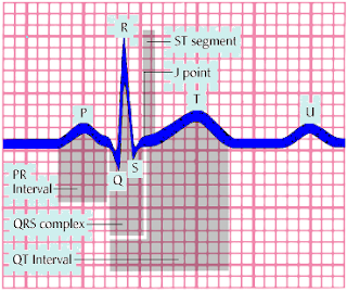
The ECG device measures and averages the differences between the electrical potential of the electrode sites for each lead and graphs them over time. This creates the standard ECG complex, called PQRST. The P wave represents atrial depolarization; the QRS complex, ventricular depolarization; and the T wave, ventricular repolarization. (See Reviewing ECG waveforms and components.)
Variations of standard ECG include exercise ECG (stress ECG) and ambulatory ECG (Holter monitoring). Exercise ECG monitors heart rate, blood pressure, and ECG waveforms as the patient walks on a treadmill or pedals a stationary bicycle. For ambulatory ECG, the patient wears a portable Holter monitor to record heart activity continually over 24 hours.
Today, ECG is typically accomplished using a multichannel method. All electrodes are attached to the patient at once, and the machine prints a simultaneous view of all leads.
Equipment
ECG machine ; recording paper ; disposable pregelled electrodes ; 4″ × 4″ gauze pads ; optional: clippers, marking pen.
Preparation of equipment
Place the ECG machine close to the patient's bed, and plug the power cord into the wall outlet. If the patient is already connected to a cardiac monitor, remove the electrodes to accommodate the precordial leads and minimize electrical interference on the ECG tracing. Keep the patient away from objects that might cause electrical interference, such as equipment, fixtures, and power cords.
Read More