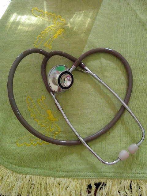
A stethoscope is a medical device for listening to sounds inside the body. The initial stethoscope was invented in the early 19th century by French physician Ren� Laennec, but was actually trying to achieve a rather different end: doctor-patient distance....
Showing posts with label equipment. Show all posts
Showing posts with label equipment. Show all posts
Tuesday, June 10, 2014
Fecal Incontinence Management
Posted by
Channel Maymoon
Labels:
documentation,
equipment,
Geriatric,
Geriatric Care,
incontinence management,
nursing procedures
at
1:56 PM
Fecal incontinence, the involuntary passage of feces, may occur
gradually (as in dementia) or suddenly (as in spinal cord injury). It usually
results from fecal stasis and impaction secondary to reduced activity,
inappropriate diet, or untreated painful anal conditions. It can also result
from chronic laxative use; reduced fluid intake; neurologic deficit; pelvic,
prostatic, or rectal surgery; and the use of certain medications, including
antihistamines, psychotropics, and iron preparations. Not usually a sign of
serious illness, fecal incontinence can seriously impair an elderly patient's
physical and psychological well-being.
Patients with urinary or fecal incontinence should be carefully
assessed for underlying disorders. Most can be treated; some can even be cured.
Treatment aims to control the condition through bladder or bowel retraining or
other behavior management techniques, diet modification, drug therapy,
pessaries, and, possibly, surgery. Corrective surgery for urinary incontinence
includes transurethral resection of the prostate in men, urethral collagen
injections for men or women, repair of the anterior vaginal wall or retropelvic
suspension of the bladder in women, urethral sling, and bladder augmentation.
Equipment
Bladder retraining record sheet • gloves • stethoscope (to
assess bowel sounds) • lubricant • moisture barrier cream • antidiarrheal
or laxative suppository • incontinence pads • bedpan • specimen container
• label • laboratory request form • optional: stool collection kit,
urinary catheter.
Implementation
Whether the patient reports urinary or fecal incontinence or both,
you'll need to perform initial and continuing assessments to plan effective
interventions.
For fecal incontinence
-
Ask the patient with fecal incontinence to identify its onset, duration, severity, and pattern (for instance, determine whether it occurs at night or with diarrhea). Focus the history on GI, neurologic, and psychological disorders.
-
Note the frequency, consistency, and volume of stools passed in the past 24 hours. Obtain a stool specimen if ordered. Protect the patient's bed with an incontinence pad.
-
Assess the patient's medication regimen. Check for drugs that affect bowel activity, such as aspirin, some anticholinergic antiparkinsonian agents, aluminum hydroxide, calcium carbonate antacids, diuretics, iron preparations, opiates, tranquilizers, tricyclic antidepressants, and phenothiazines.
-
For the neurologically capable patient with chronic incontinence, provide bowel retraining.
-
Advise the patient to consume a fiber-rich diet that includes lots of raw, leafy vegetables (such as carrots and lettuce), unpeeled fruits (such as apples), and whole grains (such as wheat or rye breads and cereals). If the patient has a lactase deficiency, suggest that he take calcium supplements to replace calcium lost by eliminating dairy products from the diet.
-
Encourage adequate fluid intake.
-
Teach the elderly patient to gradually eliminate laxative use. Point out that using laxatives to promote regular bowel movement may have the opposite effect, producing either constipation or incontinence over time. Suggest natural laxatives, such as prunes and prune juice, instead.
-
Promote regular exercise by explaining how it helps to regulate bowel motility. Even a nonambulatory patient can perform some exercises while sitting or lying in bed.
Special considerations
-
For fecal incontinence, maintain effective hygienic care to increase the patient's comfort and prevent skin breakdown and infection. Clean the perineal area frequently, and apply a moisture barrier cream. Control foul odors as well.
-
Schedule extra time to provide encouragement and support for the patient, who may feel shame, embarrassment, and powerlessness from loss of control.
Complications
Skin breakdown and infection may result from incontinence.
Psychological problems resulting from incontinence include social isolation,
loss of independence, lowered self-esteem, and depression.
Documentation
Record all bladder and bowel retraining efforts, noting scheduled
bathroom times, food and fluid intake, and elimination amounts, as appropriate.
Document the duration of continent periods. Note any complications, including
emotional problems and signs of skin breakdown and infection as well as the
treatments given for them.
Monday, June 9, 2014
Supplies and Use of Supplies for Phlebotomy Venipuncture Procedure
Posted by
Channel Maymoon
Labels:
equipment,
nursing procedures,
nursing skills,
Phlebotomy,
procedures,
techniques
at
2:42 PM
By establishing a procedure for the correct collection of blood by venipuncture many pre-
analytical errors and patient management complications can be avoided. Patient safety is the ultimate goal above all other considerations. Cost, efficiency, etc are secondary to ensuring that in no way will the patient be harmed by the phlebotomy procedure. This includes all aspects of the procedure including ordering, drawing, labeling, handling and transporting the specimen. The quality of the patient results is directly dependent upon the quality of the specimen. By providing the highest standard of safety and quality of care customer service satisfaction can be achieved.
Supplies and use of supplies –(Refer to Standard Phlebotomy Tray policy)
1. Blood Collecting Trays
- Blood collecting trays should be lightweight and easy to handle with enough space and compartments for the various supplies.
2. Gloves
- Disposable latex, vinyl, polyethylene, or nitrile gloves provide barrier protection and must be worn for all venipuncture procedures to comply with OSHA regulations.
Latex free gloves must be worn for all patients with a hypersensitivity to latex proteins.
3. Hubs
–
a. All Vacutainer holders are to be SINGLE USE.
OSHA states “Blood tube holders, with needle attached, must be immediately discarded into an accessible sharps container after safety feature has been activated”. The re-use of vacutainer blood tube holders is strictly prohibited by OSHA and BVHS. (According to OSHA, “removing contaminated needles and re-using blood tube holders can expose workers to multiple hazards.”
b. Specimen transfer hubs are also available and, for our safety, are to be used before attempting to use a transfer needle. To use this device, simply attach it to the syringe and place the/each necessary vacutainer tube in the in the vacutainer holder until the appropriate amount of specimen is transferred.
4. Needles
- A large gauge (G) number indicates a small needle, while a small gauge number indicates alarge needle.
Needles must always be sterile and should never be recapped.
In order to prevent potential worker exposure, the needle safety feature should be activated immediately after specimen collection and discarded without disassembly into a sharps container. Needles are single use only.
a. BD Hypodermic needles
b. Butterfly “Push Button”
5. Sterile Syringes
-Sterile syringes must remain sterile. If removed from their container and not used immediately they are no longer considered sterile and are not to be used.
6. Blood Collection Tubes
- Venous blood collection tubes are manufactured to withdraw a predetermined volume of blood.
7. Tourniquets
-Tourniquets must be discarded immediately when contamination with blood or body fluids is obvious or suspected. Before drawing any in-patients, be sure to look around the room,
typically next to the sharps container, for a tourniquet that is specific for that patient. Out-patient draws and off-site tourniquets are replaced daily, or upon any sign of obvious or suspectedcontamination.
8. Antiseptics
- 70% isopropyl, PVP iodine prep pads, or 2% Iodine Tincture.
9. Gauze Pads
-Small, gauze pads should be available. Cotton balls may also be used.
10. Puncture-Resistant Disposable Container
-An approved puncture-resistant disposable container that is compliant with national or local regulations must be available for the disposal of the contaminated needle assembly. Such containers typically have a color regulated by each country, and a biohazard symbol.
11. Ice
-Ice or refrigerant should be available for specimens that require immediate chilling.
12. Bandages/Tape
- Adhesive bandages, preferably hypoallergenic, should be available, as well as gauze wraps for sensitive or fragile skin.
13. Warming Devices
- Warming devices may be used to dilate blood vessels and increase flow. When using commercial warmers follow the manufacturer’s recommendation. Warming devices should not exceed 42º C.
14. Specimen Collection Manual/Reference Lab Manual
- A test manual listing the tube(s) and volume requirements for various tests, specimen handling instructions, and precautions is available on all computers.
Friday, October 11, 2013
ECG Waveforms And Components
The electrocardiogram (ECG) is a graphic recording ofelectric potentials generated by the heart.The signals are detected by means of metal electrodes attached to the extremities and chest wall and are then amplified and recorded by the electrocardiograph. ECG leads actually display the instantaneous differences in potential between these electrodes.
The clinical utility of the ECG derives from its immediate availability as a noninvasive, inexpensive, and highly versatile test. In addition to its use in detecting arrhythmias, conduction disturbances, and myocardial ischemia, electrocardiography may reveal other findings related to life-threatening metabolic disturbances (e.g., hyperkalemia) or increased susceptibility to sudden cardiac death (e.g., QT prolongation syndromes). The widespread use of coronary fibrinolysis and acute percutaneous coronary interventions in the early therapy of acute myocardial infarction has refocused attention on the sensitivity and specificity of ECG signs of myocardial ischemia.
An electrocardiogram (ECG) waveform has three basic components: the P wave, QRS complex, and T wave. These elements can be further divided into the PR interval, J point, ST segment, U wave, and QT interval.
P wave and PR interval
The P wave represents atrial depolarization. The PR interval represents the time it takes an impulse to travel from the atria through the atrioventricular nodes and bundle of His. The PR interval measures from the beginning of the P wave to the beginning of the QRS complex.
QRS complex
The QRS complex represents ventricular depolarization (the time it takes for the impulse to travel through the bundle branches to the Purkinje fibers).
The Q wave appears as the first negative deflection in the QRS complex; the R wave, as the first positive deflection. The S wave appears as the second negative deflection or the first negative deflection after the R wave.
J point and ST segment
Marking the end of the QRS complex, the J point also indicates the beginning of the ST segment. The ST segment represents part of ventricular repolarization.
T wave and U wave
Usually following the same deflection pattern as the P wave, the T wave represents ventricular repolarization. The U wave follows the T wave, but isn't always seen.
QT interval
The QT interval represents ventricular depolarization and repolarization. It extends from the beginning of the QRS complex to the end of the T wave.
Wednesday, August 28, 2013
Electrocardiography: Equipment Preparation
Posted by
Channel Maymoon
Labels:
Cardiovascular Care,
ECGelectrocardiography,
electrocardiography,
equipment,
nursing,
preparation,
procedures
at
1:57 PM
One of the most valuable and frequently used diagnostic tools, electrocardiography (ECG) measures the heart's electrical activity as waveforms. Impulses moving through the heart's conduction system create electric currents that can be monitored on the body's surface. Electrodes attached to the skin can detect these electric currents and transmit them to an instrument that produces a record (the electrocardiogram) of cardiac activity.
ECG can be used to identify myocardial ischemia and infarction, rhythm and conduction disturbances, chamber enlargement, electrolyte imbalances, and drug toxicity.
The standard 12-lead ECG uses a series of electrodes placed on the extremities and the chest wall to assess the heart from 12 different views (leads). The 12 leads consist of three standard bipolar limb leads (designated I, II, III), three unipolar augmented leads (aVR, aVL, aVF), and six unipolar precordial leads (V1 to V6). The limb leads and augmented leads show the heart from the frontal plane. The precordial leads show the heart from the horizontal plane.
The ECG device measures and averages the differences between the electrical potential of the electrode sites for each lead and graphs them over time. This creates the standard ECG complex, called PQRST. The P wave represents atrial depolarization; the QRS complex, ventricular depolarization; and the T wave, ventricular repolarization. (See Reviewing ECG waveforms and components.)
Variations of standard ECG include exercise ECG (stress ECG) and ambulatory ECG (Holter monitoring). Exercise ECG monitors heart rate, blood pressure, and ECG waveforms as the patient walks on a treadmill or pedals a stationary bicycle. For ambulatory ECG, the patient wears a portable Holter monitor to record heart activity continually over 24 hours.
Today, ECG is typically accomplished using a multichannel method. All electrodes are attached to the patient at once, and the machine prints a simultaneous view of all leads.
Today, ECG is typically accomplished using a multichannel method. All electrodes are attached to the patient at once, and the machine prints a simultaneous view of all leads.
Equipment
ECG machine ; recording paper ; disposable pregelled electrodes ; 4″ × 4″ gauze pads ; optional: clippers, marking pen.
ECG machine ; recording paper ; disposable pregelled electrodes ; 4″ × 4″ gauze pads ; optional: clippers, marking pen.
Preparation of equipment
Place the ECG machine close to the patient's bed, and plug the power cord into the wall outlet. If the patient is already connected to a cardiac monitor, remove the electrodes to accommodate the precordial leads and minimize electrical interference on the ECG tracing. Keep the patient away from objects that might cause electrical interference, such as equipment, fixtures, and power cords.
Subscribe to:
Comments (Atom)
Powered by Blogger.


